


 37 / 76

CARDIOVASCULAR JOURNAL OF AFRICA • Volume 29, No 4, July/August 2018
AFRICA
235
reduction in global longitudinal strain.
22
This compensatory increase in twist present in the CKD
patient was documented in a study by Panoulas
et al
.
19
This
study demonstrated that twist increases while longitudinal
strain decreases in CKD patients with preserved EF. This is
postulated to represent an adaptive mechanism to preserve
EF in the face of declining longitudinal myocardial function.
The increase in twist was inversely related to worsening GFR
in patients with preserved EF.
19
By contrast, our study, which
only included very late-stage CKD patients, showed significant
decrease in the apical myocardial rotation with no difference in
net twist found in haemodialysis patients compared to controls.
This is despite there being no difference in baseline EF between
the dialysis patients and controls, implying that diminution of
apical rotation and a normal LV twist as opposed to an expected
increase in twist may be an early indicator of further myocardial
dysfunction and loss of compensatory mechanisms aimed to
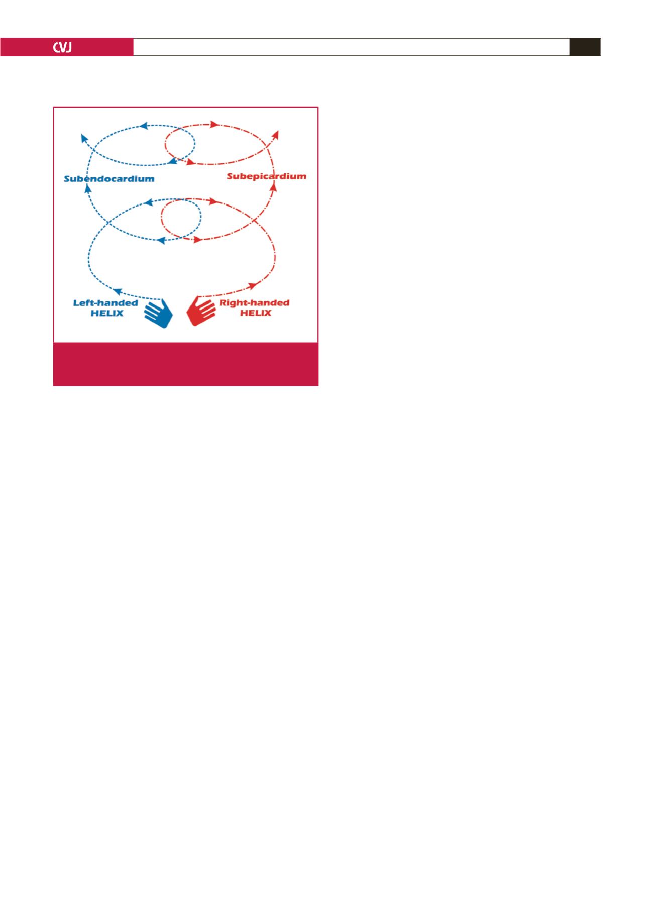
preserve EF (Fig. 4).
The limitations of this study are that it was a pilot study using
a single vendor (Philips Healthcare). The small sample size was
due to the size of the haemodialysis patient population at our
institution. This did not allow adequate numbers to perform
multiple linear regression analysis. The homogenous nature of
our study population may not translate to other patient cohorts.
It would be useful in larger studies to determine how twist
is affected in CKD patients with and without hypertension.
Multicentre studies with longitudinal follow up may confirm the
findings of this study.
Conclusion
LV twist and its derived rotational parameters did not change
significantly post-dialysis compared to pre-dialysis. This may
suggest that these parameters are less affected by varying loading
conditions post-dialysis. The decrease in apical rotation observed
in late-stage CKD patients compared to controls may represent
an early marker of loss of rotational compensation, which
preserves EF in the CKD patient
This study was supported by unrestricted research grants by Medtronic Ltd,
Servier Ltd and Novartis AG.
References
1.
Foley RN, Parfrey PS, Sarnak MJ. Epidemiology of cardiovascular
disease in chronic renal disease.
J Am Soc Nephrol
1998;
9
(12 Suppl):
S16–23.
2.
Herzog CA, Asinger RW, Berger AK, Charytan DM, Diez J, Hart RG,
et al
. Cardiovascular disease in chronic kidney disease. A clinical update
from Kidney Disease: Improving Global Outcomes (KDIGO).
Kidney
Int
2011;
80
(6): 572–586.
3.
Herzog CA, Mangrum JM, Passman R. Sudden cardiac death and
dialysis patients.
Semin Dial
2008;
21
(4): 300–307.
4.
McIntyre CW. Effects of hemodialysis on cardiac function.
Kidney Int
2009;
76
(4): 371–375.
5.
Burton JO, Korsheed S, Grundy BJ, McIntyre CW. Hemodialysis-
induced left ventricular dysfunction is associated with an increase in
ventricular arrhythmias.
Ren Fail
2008;
30
(7): 701–709.
6.
Burton JO, Jefferies HJ, Selby NM, McIntyre CW. Hemodialysis-
induced cardiac injury: determinants and associated outcomes.
Clin J
Am Soc Nephrol
2009;
4
(5): 914–920.
7.
Nixon JV, Mitchell JH, McPhaul JJ, Jr, Henrich WL. Effect of hemo-
dialysis on left ventricular function. Dissociation of changes in filling
volume and in contractile state.
J Clin Invest
1983;
71
(2): 377–384.
8.
Perk G, Tunick PA, Kronzon I. Non-Doppler two-dimensional strain
imaging by echocardiography – from technical considerations to clinical
applications.
J Am Soc Echocardiogr
2007;
20
(3): 234–243.
9.
Helle-Valle T, Crosby J, Edvardsen T, Lyseggen E, Amundsen BH,
Smith HJ,
et al
. New noninvasive method for assessment of left ventricu-
lar rotation: speckle tracking echocardiography.
Circulation
2005;
112
(20): 3149–3156.
10. Yan P, Li H, Hao C, Shi H, Gu Y, Huang G,
et al.
2D-speckle tracking
echocardiography contributes to early identification of impaired left
ventricular myocardial function in patients with chronic kidney disease.
Nephron Clin Pract
2011;
118
(3): c232–240.
11. Choi JO, Shin DH, Cho SW, Song YB, Kim JH, Kim YG,
et al
. Effect
of preload on left ventricular longitudinal strain by 2D speckle tracking.
Echocardiography
2008;
25
(8): 873–879.
12. Murata T, Dohi K, Onishi K, Sugiura E, Fujimoto N, Ichikawa K,
et al.
Role of haemodialytic therapy on left ventricular mechanical
dyssynchrony in patients with end-stage renal disease quantified by
speckle-tracking strain imaging.
Nephrol Dial Transplant
2011;
26
(5):
1655–1661.
13. Nakatani S. Left ventricular rotation and twist: why should we learn?
J
Cardiovasc Ultrasound
2011;
19
(1): 1–6.
14. Hansen DE, Daughters GT, 2nd, Alderman EL, Ingels NB, Jr., Miller
DC. Torsional deformation of the left ventricular midwall in human
hearts with intramyocardial markers: regional heterogeneity and sensi-
tivity to the inotropic effects of abrupt rate changes.
Circ Res
1988;
62
(5): 941–952.
15. Buchalter MB, Rademakers FE, Weiss JL, Rogers WJ, Weisfeldt ML,
Shapiro EP. Rotational deformation of the canine left ventricle meas-
ured by magnetic resonance tagging: effects of catecholamines, ischae-
mia, and pacing.
Cardiovasc Res
1994;
28
(5): 629–635.
16. Ahmed MI, Desai RV, Gaddam KK, Venkatesh BA, Agarwal S, Inusah
S,
et al
. Relation of torsion and myocardial strains to LV ejection frac-
Fig 4.
Myocardial fibre orientation and direction. Left-handed
helical orientation of the sub-endocardium. Right-
handed helical arrangement of the sub-epicardium.