


 10 / 62

CARDIOVASCULAR JOURNAL OF AFRICA • Volume 31, No 4, July/August 2020
172
AFRICA
carnitine/malate, respectively, as substrates. The effects of
ischaemia and reperfusion follow a very similar pattern on
mitochondrial oxygen uptake state 3 and the ox-phos rate
(ADP/O ratio
×
QO
2
state 3). With both substrates, exposure
of the isolated heart to 25 or 30 minutes of global ischaemia
resulted in a reduction in mitochondrial oxygen uptake as well as
ox-phos rate (state 3) compared to stabilisation; with glutamate/
malate as substrates, the change became significant after 30
minutes only, whereas with palmitoyl-L carnitine/malate the
reduction in QO
2
(state 3) and ox-phos rate was significant after
both 25 and 30 minutes of ischaemia.
There was a tendency for these two parameters to increase
with reperfusion: with glutamate/malate, the increases observed
after both 25 and 30 minutes ischaemia/reperfusion were not
significant. With palmitoyl-L carnitine/malate as substrate,
the increase in QO
2
(state 3) (but not in ox-phos rate) after 25
minutes of ischaemia/reperfusion was significant.
Interestingly, with glutamate as substrate, exposure of the
hearts to 25 or 30 minutes of ischaemia with or without
reperfusion had no significant effects on state 4 respiration. On
the other hand with palmitoyl-L carnitine as substrate, QO
2
state
(state 4) was significantly reduced by ischaemia, and increased by
reperfusion after 25 minutes of ischaemia.
With glutamate/malate as substrates, a reduction in the RCI
values after 25 and 30 minutes of ischaemia and an increase
after reperfusion, respectively, were observed. Similar tendencies
were observed when palmitoyl-L carnitine/malate were used as
substrates.
Pre-treatment with chloroquine had the most marked effects
on mitochondrial function of hearts exposed to 30 minutes of
ischaemia. With both substrates, pre-treatment with chloroquine
caused a significant increase in QO
2
, states 3 and 4. Similarly,
chloroquine treatment prior to exposure to 25 or 30 minutes of
ischaemia increased the ox-phos rate. With glutamate/malate as
well as palmitoyl carnitine/malate as substrates, the changes were
significant after 30 minutes of ischaemia, while the chloroquine-
induced increases seen after reperfusion were not significant.
In accordance with its effects on QO
2
(state 3), the RCI values
obtained after 30 minutes of ischaemia were significantly
increased when incubated with both substrate combinations.
The ability of mitochondria isolated after stabilisation,
ischaemia or reperfusion to withstand oxidative stress was further
evaluated by exposing the mitochondria in the oxygraph chamber
to anoxia followed by re-oxygenation (Figs 3 and 4). Interestingly,
with both substrates, mitochondria isolated after the stabilisation
phase showed an increase in the percentage state 3 recovery,
compared with the values obtained before exposure to anoxia,
while mitochondria isolated after exposure to ischaemia with or
without reperfusion showed an almost 100% recovery in QO
2
state
3, with no differences between the groups. Similar tendencies were
observed in the chloroquine-treated hearts, the only difference
being that chloroquine treatment caused an increase in state 3
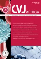
Aortic flow
Group
CON
CQ
50
40
30
20
10
0
ml/min
–1
*
HR
Group
CON
CQ
300
200
100
0
Beats/min
W total
Group
CON
CQ
15
10
5
0
mW
*
Cardiac output
Group
CON
CQ
60
40
20
0
ml/min
*
Fig. 2.
Baseline function of working rat hearts during stabilisation: effect of chloroquine pre-treatment (
n
= 10 hearts/group). CON:
control; CQ: chloroquine pre-treatment (10 mg/kg); HR: heart rate (beats/min); W total: work total (mW).