


 11 / 62

CARDIOVASCULAR JOURNAL OF AFRICA • Volume 31, No 4, July/August 2020
AFRICA
173
QO
2
(S3)
AMC
STB
25
′
ISC
30
′
ISC
25
′
ISC + RP
30
′
ISC + RP
STB
25
′
ISC
30
′
ISC
25
′
ISC + RP
30
′
ISC + RP
Con
Con+CQ
400
300
200
100
0
nAtoms O/mg prot/min
*
p
≤
0.023
QO
2
(S4)
AMC
STB
25
′
ISC
30
′
ISC
25
′
ISC + RP
30
′
ISC + RP
STB
25
′
ISC
30
′
ISC
25
′
ISC + RP
30
′
ISC + RP
Con
Con+CQ
60
40
20
0
nAtoms O/mg prot/min
*
p
≤
0.01
p
≤
0.03
p
≤
0.0004
RCI
AMC
STB
25
′
ISC
30
′
ISC
25
′
ISC + RP
30
′
ISC + RP
STB
25
′
ISC
30
′
ISC
25
′
ISC + RP
30
′
ISC + RP
Con
Con+CQ
10
8
6
4
2
0
Ratio (S3/S4)
*
p
≤
0.002
p
≤
0.04
p
≤
0.001
Ox-phos rate (S3)
AMC
STB
25
′
ISC
30
′
ISC
25
′
ISC + RP
30
′
ISC + RP
STB
25
′
ISC
30
′
ISC
25
′
ISC + RP
30
′
ISC + RP
Con
Con+CQ
600
400
200
0
Ratio (S3/S4)
*
*
p
≤
0.045
p
≤
0.05
p
≤
0.004
p
≤
0.45
*
Recovery after re-oxygenation
AMC
STB
25
′
ISC
30
′
ISC
25
′
ISC + RP
30
′
ISC + RP
STB
25
′
ISC
30
′
ISC
25
′
ISC + RP
30
′
ISC + RP
Con
Con+CQ
200
150
100
50
0
Recovery in %
*
*
p
≤
0.0009
p
≤
0.35
p
≤
0.03
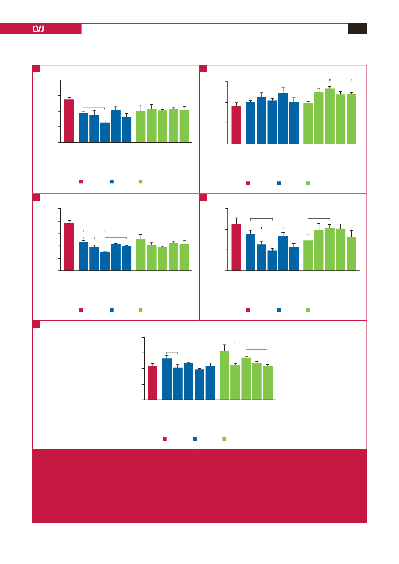
Fig. 3.
Effects of ischaemia/reperfusion and chloroquine pre-treatment on mitochondrial function with glutamate/malate as
substrates (
n
= 5 hearts/group). Measurements of mitochondrial function were made after 40 minutes of stabilisation; after
25 minutes of global ischaemia; after 10 minutes of reperfusion following 25 minutes of global ischaemia; after 30 minutes of
global ischaemia; after 10 minutes of reperfusion following 30 minutes of global ischaemia. Mitochondria were also prepared
from hearts of age-matched control rats for comparison purposes. A. QO
2
(state 3) (nAtoms oxygen/mg protein/min); B. QO
2
(state 4) (nAtoms oxygen/mg protein/min); C. RCI (state 3/state 4); D. ox-phos rate (nmoles ATP/mg prot/min); E. percentage
recovery after re-oxygenation. *
p
≤
0.05 vs corresponding untreated control rats. AMC: age-matched control; CON: control;
CQ: chloroquine; STB: stabilisation; ISC: ischaemia; RP: reperfusion; ox-phos: oxidative phosphorylation; RCI: respiratory
control index.
A
C
E
B
D