CARDIOVASCULAR JOURNAL OF AFRICA • Vol 21, No 1, January/February 2010
AFRICA
33
in African-American children, but they represented only 11% of
the patients studied.
Little has been published regarding VCFS in South African
children, especially among the indigenous population. The aim
of this study was to determine the cardiac abnormalities as well
as selected facial anthropometric measurements in affected chil-
dren from the Free State and Northern Cape.
Methods
The study was a prospective, descriptive investigation of children
presenting at the Cardiology Unit of the Universitas Academic
Hospital complex. All patients with one or more facial features
suggestive of 22q11 microdeletion were included in this study.
A positive fluorescence
in situ
hybridisation (FISH) analysis was
required as proof of a microdeletion. The Vysis
®
LSI TUPLE 1
probe set containing the LSI TUPLE1 probe for chromosomal
regions TUPLE1, D22S55, D22S609 and D22S942 with a LSI
ARSA control probe was used (supplied by The Scientific
Group, Johannesburg, SA).
Patient evaluation included a clinical examination as well as a
routine complete paediatric echocardiogram. Echocardiography
was performed using a Philips 5500 apparatus and appropriate
transducers, using standard views. A cardiologist reviewed all
the echocardiograms. Follow-up data were obtained from clini-
cal records.
Seventeen pre-selected standard craniofacial anthropometric
measurements were performed using digital sliding callipers
where possible. This leg of the study was started three years after
the commencement of the initial trial. All measurements were
performed as described by Farkas.
11
One of the authors took all
the measurements. For a more detailed description of the anthro-
pometric measurements, see Fig. 2.
Both the initial and subsequent protocols were approved
by the Ethics Committee of the Faculty of Health Sciences,
University of the Free State (ETOVS 118/99). Written informed
consent was obtained from the parent or legal guardian of the
patient and verbal consent from the children as far as possible.
Statistical analysis
Data were captured using Microsoft Excel spreadsheets and
statistical analyses were performed by the Department of
Biostatistics, University of the Free State, as well as with a
commercially available software package, GraphPad Prism
version 5.00 (GraphPad software, San Diego, California, USA).
A
p
-value less than 0.05 was considered statistically significant,
while 95% confidence intervals (CI) were used where clinically
indicated.
Z
-values were obtained using a standard formula to
compare anthropometric measurements with reference to a
standard set of normal values.
11
Results
A total of 334 FISH analyses were done over an eight-year period
(1999–2007), resulting in 40 patients being identified with the
microdeletion. The median age at diagnosis (positive FISH
probe) was 3.6 years with a range of 0.04 years (two weeks to
16.2 years). Twenty-one (52.5%) of the patients were male. The
group consisted of 23 African and 10 Caucasian children, and
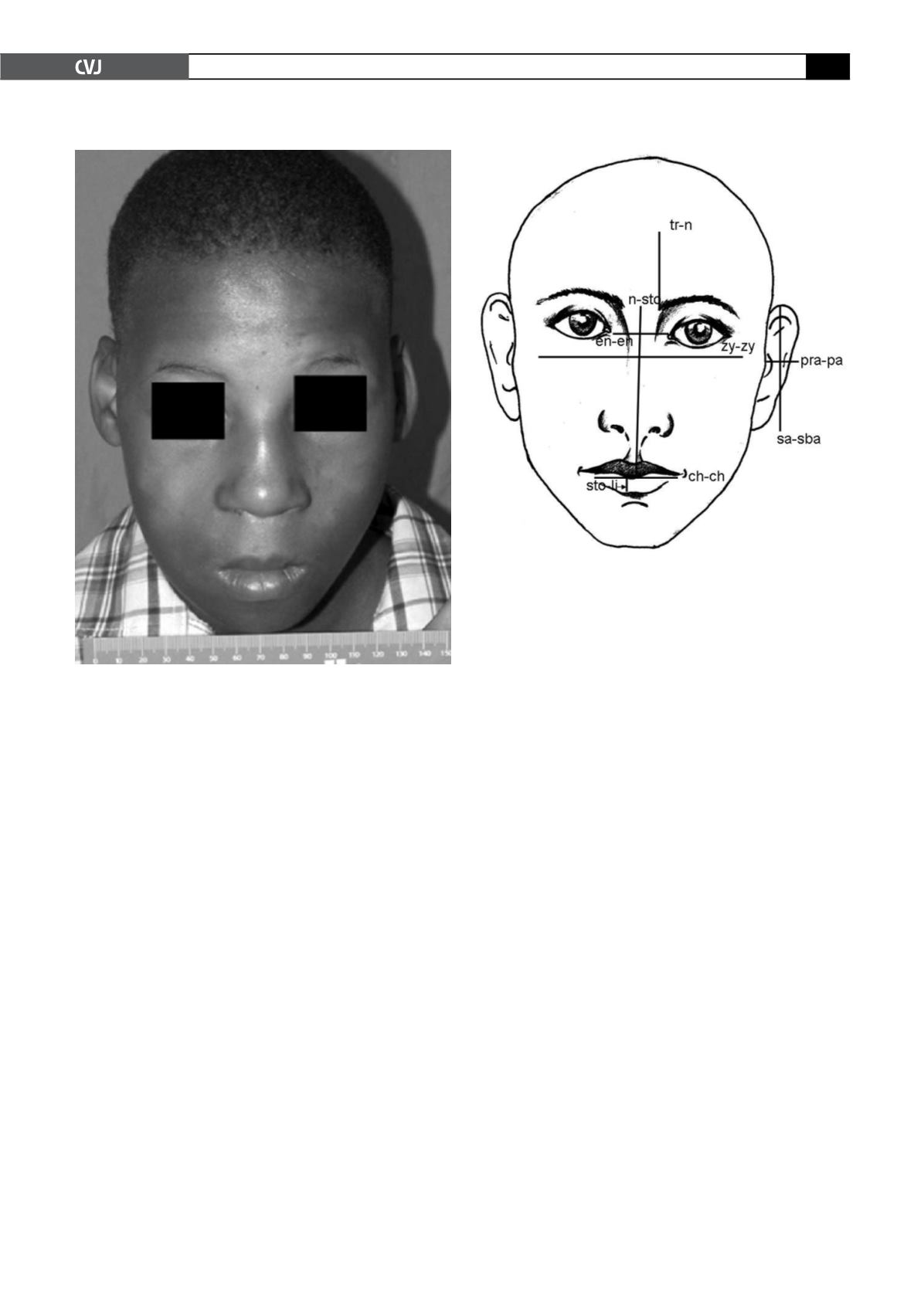
Fig. 1. Typical example of a patient with 22q11 microdele-
tion. Note the broad nasal root, and abnormal ears and
mouth.
Fig. 2. Position of some anthropometric measurements.
tr-n
=
height of forehead; n-sto
=
physiognomical height
of the upper face; en-en
=
intercanthal width; zy-zy
=
width of face; sto-li
=
vermilion height of lower lip; ch-ch
=
width of mouth; pra-pa
=
width of auricle; sa-sba
=
length
of auricle.


