CARDIOVASCULAR JOURNAL OF AFRICA • Vol 24, No 4, May 2013
134
AFRICA
peripheral vasoconstriction and volume retention.
30,31
BNP is known to suppress renin release and one reason for
its activation could be the derangement in the renin angiotensin
system that occurs in PE.
31
To what extent the pro-inflammatory
cytokines and endothein-1 are known to stimulate natriuretic
peptide release is not clear, nor is their relationship with cardiac
filling pressures known.
While it appears that the increase in circulating plasma BNP
is most likely explained by the rising cardiac filling pressure, it
should be remembered that PE is documented to be a relatively
volume-contracted state with marked peripheral vasoconstriction.
In this regard, elevation in BNP levels may be due to myocardial
remodelling and sub-clinical ventricular dysfunction that
accompanies the severe vasoconstriction that is observed in PE.
Our findings suggest that BNP reflects the myocardial load in
PE. This hypothesis is confirmed by the decrease in BNP levels
in the puerperium when the placenta has been removed.
As expected, significant changeswere seen in the pre-eclamptic
group with regard to blood pressure, pedal oedema, proteinuria,
uric acid and serum creatinine levels, highlighting the need
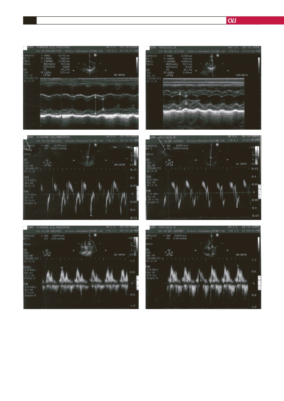
Fig. 2. M-mode Doppler and pulse-wave recordings in a normotensive patient (A) and in a pre-eclamptic patient (B).
A
B
A
A
B
B


