


 72 / 80

CARDIOVASCULAR JOURNAL OF AFRICA • Volume 28, No 3, May/June 2017
e2
AFRICA
as non-cardiac during a routine forensic autopsy. The heart
weight was 613 g.
After a month of fixation in 10% buffered formaldehyde,
the heart was examined and it revealed PLSVC drains into the
CS (Fig. 1). The mediolateral (ML) and anteroposterior (AP)
diameters of the PLSVC, measured 1 cm above its connection
with the CS, were 12.2 and 11.5 mm, respectively. The mean
thickness of the LSVC was 0.6 mm. Further examination
revealed an enormous coronary sinus with a funnel-shaped
expansion at the PLSVC orifice. The CS diameter, measured in
the middle of the structure, was greatly enlarged (15.85 mm). The
CS ostium was also enlarged, measuring 17.2 mm in diameter.
The CS ostium valve (Thebesian valve) was absent (Fig. 2).
The great cardiac vein had a relatively small ostium (diameter 2.3
mm) and lacked a Vieussens valve (Fig. 3). Other venous valves
were also absent within the ostia of the middle cardiac vein and
posterior vein of the left ventricle (diameter of veins < 1 mm).
The small cardiac vein was absent. The length of the CS, as
measured from the ostium of the great cardiac vein to the CS
orifice, was 43.7 mm. The right superior vena cava was present
with a small ostium diameter (ML = 14.3 mm; AP = 14.9 mm).
Distortions of the atrial dimensions were noted; reduction in
the AP length of the left atrium and enlargement of the right
atrium. The dimensions of the atrioventricular rings were also
measured; mitral ring (AP = 26.5 mm; ML = 12.4 mm; area = 2.6
cm
2
) and tricuspid ring (AP = 31.4 mm; ML = 21.6 mm; area = 5.3
cm
2
). The inferior vena cava ostium diameters were AP = 28.6 mm
and ML = 33.8 mm. The Eustachian valve was present (Fig. 2).
An anomaly of the pulmonary vein pattern was observed;
there was a common trunk of the left superior and left inferior
pulmonary veins (diameter 17.8 mm) and an additional middle
right pulmonary vein (diameter 2.7 mm) with two classic right
pulmonary veins (Fig. 4). The patent foramen ovale was absent
and a left-sided septal pouch was observed.
11
Fig. 5 shows how
measurements were performed.
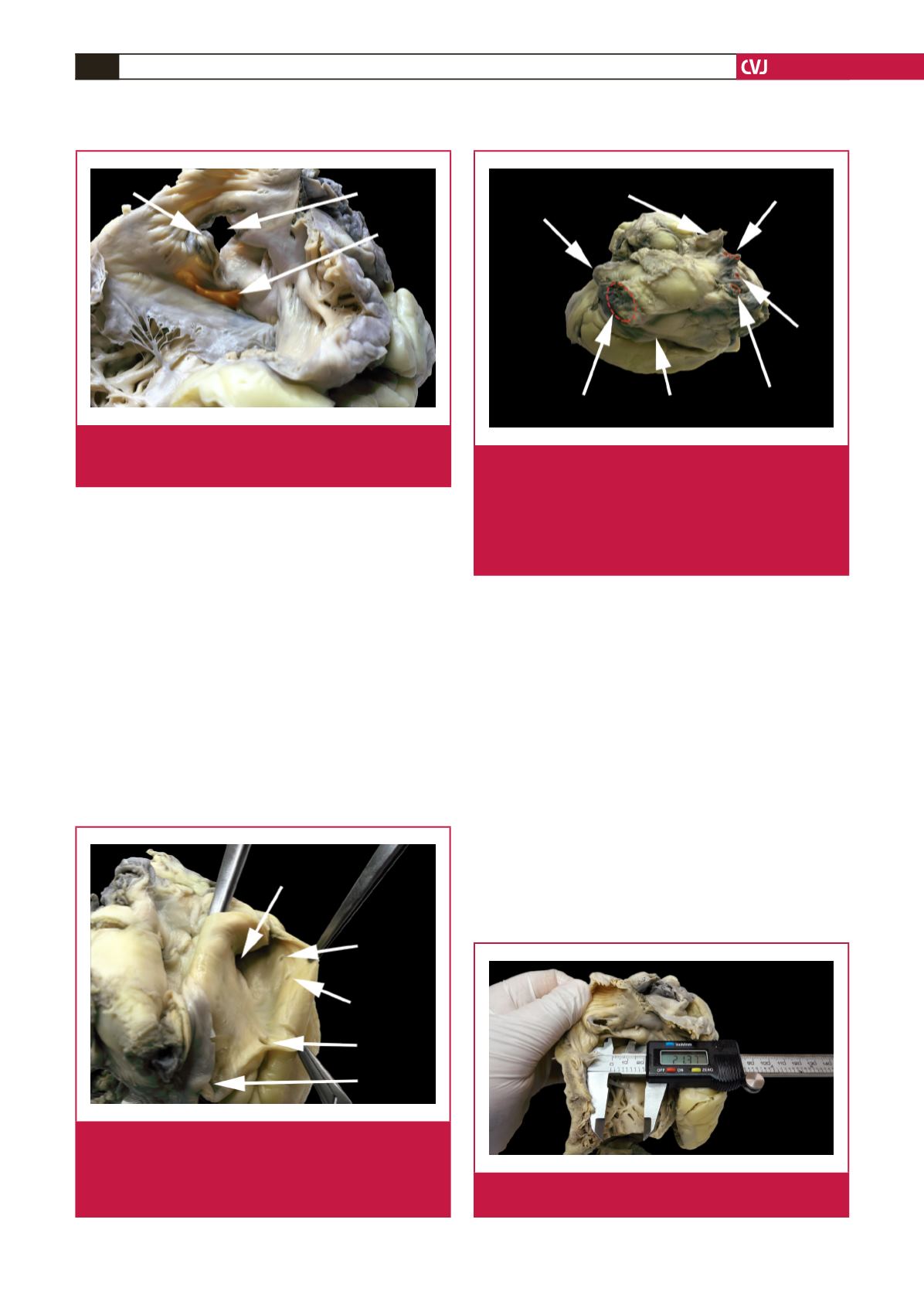
CSO
IVC
EuchV
Fig. 2.
View of the internal surface of the right atrium. CSO,
coronary sinus ostium; EuchV, Eustachian valve; IVC,
inferior vena cava.
PLSVC
GCV
PVLV
MCV
CSO
LC
Fig. 3.
View of the internal surface of the ostia of the coronary
sinus and its main tributaries. CSO, coronary sinus
ostium; GCV, great cardiac vein; MCV, middle cardiac
vein; PLSVC, persistent left superior vena cava; PVLV,
posterior vein of the left ventricle.
PLSVC
SVC
RS
RM
RI
CS
LC
Fig. 4.
View of the posterior and superior wall of the left
atrium. CS, coronary sinus; LC, common trunk of the
left superior and left inferior pulmonary veins; PLSVC,
persistent left superior vena cava; RI, right inferior
pulmonary vein; RM, right middle pulmonary vein; RS,
right superior pulmonary vein; SVC, superior vena
cava.
Fig. 5.
Measurements were performed with electronic cali-
pers with 0.01-mm precision.