


 53 / 78

CARDIOVASCULAR JOURNAL OF AFRICA • Volume 28, No 6, November/December 2017
AFRICA
391
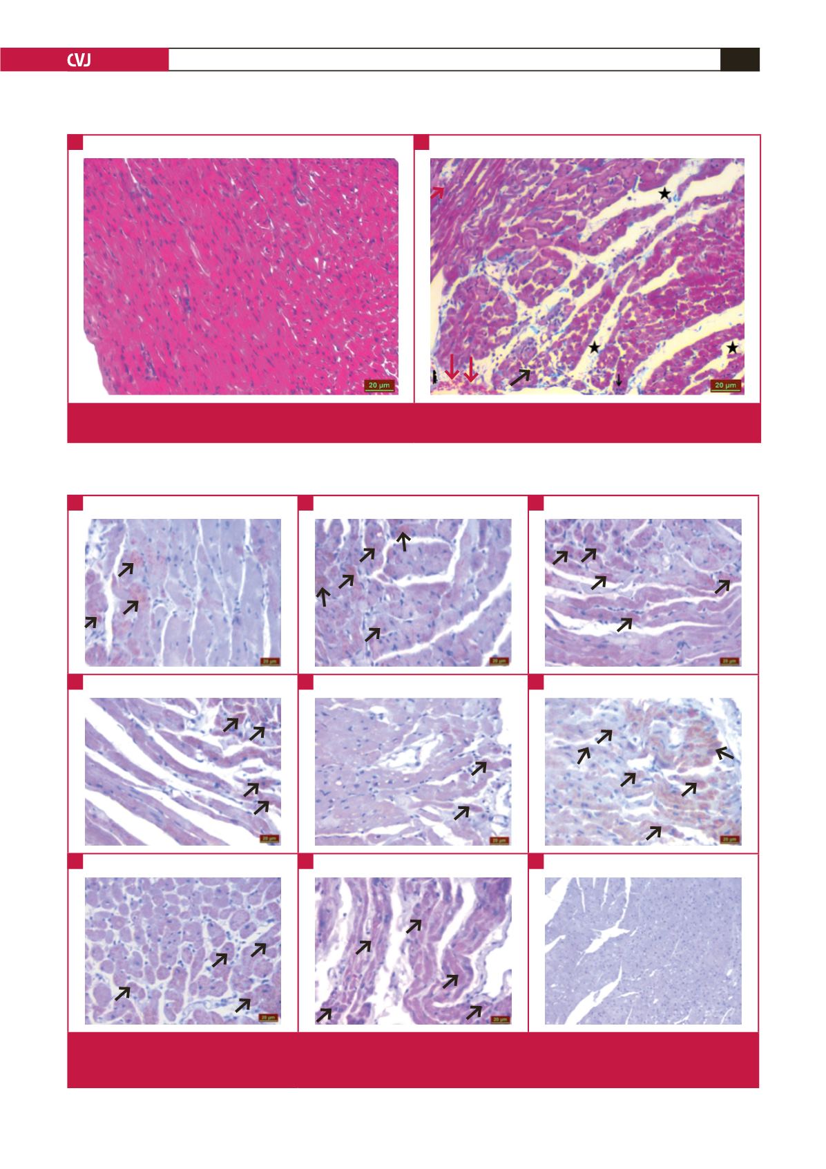
Fig. 1.
Ischaemic and control heart tissues after Masson’s trichrome staining. The ischaemic group shows an increase in inflamma-
tory cells (black arrow), congestion (red arrow), impairment of tissue integrity and oedema (black asterisk).
A
B
Fig. 2.
Irisin immunoreactivity in the heart tissues with iloprost (ILO) and sildenafil (SIL) administration in cardiac ischaemia (MI)
induced by left coronary artery ligation. Control (A), ILO (B), SIL (C), ILO
+
SIL (D), MI
+
ILO (F), MI
+
SIL (G), MI
+
ILO
+
SIL (H), and negative control; no irisin immunoreactivity (I).
A
D
G
B
E
H
C
F
I