


 58 / 62

CARDIOVASCULAR JOURNAL OF AFRICA • Volume 31, No 1, January/February 2020
e2
AFRICA
Angiographically, coronary artery aneurysms and stenosis are
the most frequently detected lesions. Local coronary vasculitis
causing fibrous intimal thickening may result in coronary
occlusion with subsequent development of acute coronary
syndromes. The left anterior descending coronary artery is
affected in most cases and aneurysm rupture is the most common
Fig. 3.
Coronary computed tomography scan images. A shows a giant aneurysm of the left anterior descending artery (arrowhead).
B shows the left anterior descending artery aneurysm compressing the pulmonary trunk (arrowhead). C shows a small
aneurysm of the distal right coronary artery (arrowhead).
A
B
C
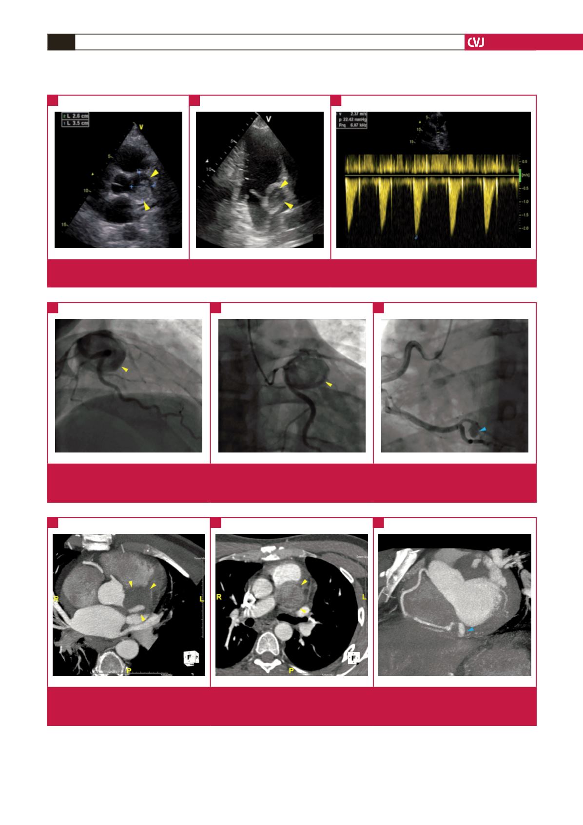
Fig. 2.
Coronary angiogram images. A and B show severe stenosis of the left main artery and a giant aneurysm (arrowhead) of
the proximal left anterior descending artery with a slow TIMI flow. C shows a small aneurysm (arrowhead) of the distal right
coronary artery.
A
B
C
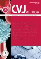
Fig. 1.
Echocardiographic images. A and B show the giant aneurysm of the left anterior descending artery (arrowhead). C shows
accelerated flow inside the aneurysm by continuous-wave Doppler.
A
B
C