CARDIOVASCULAR JOURNAL OF AFRICA • Vol 24, No 8, September 2013
AFRICA
327
William Nelson ECG quiz
Question
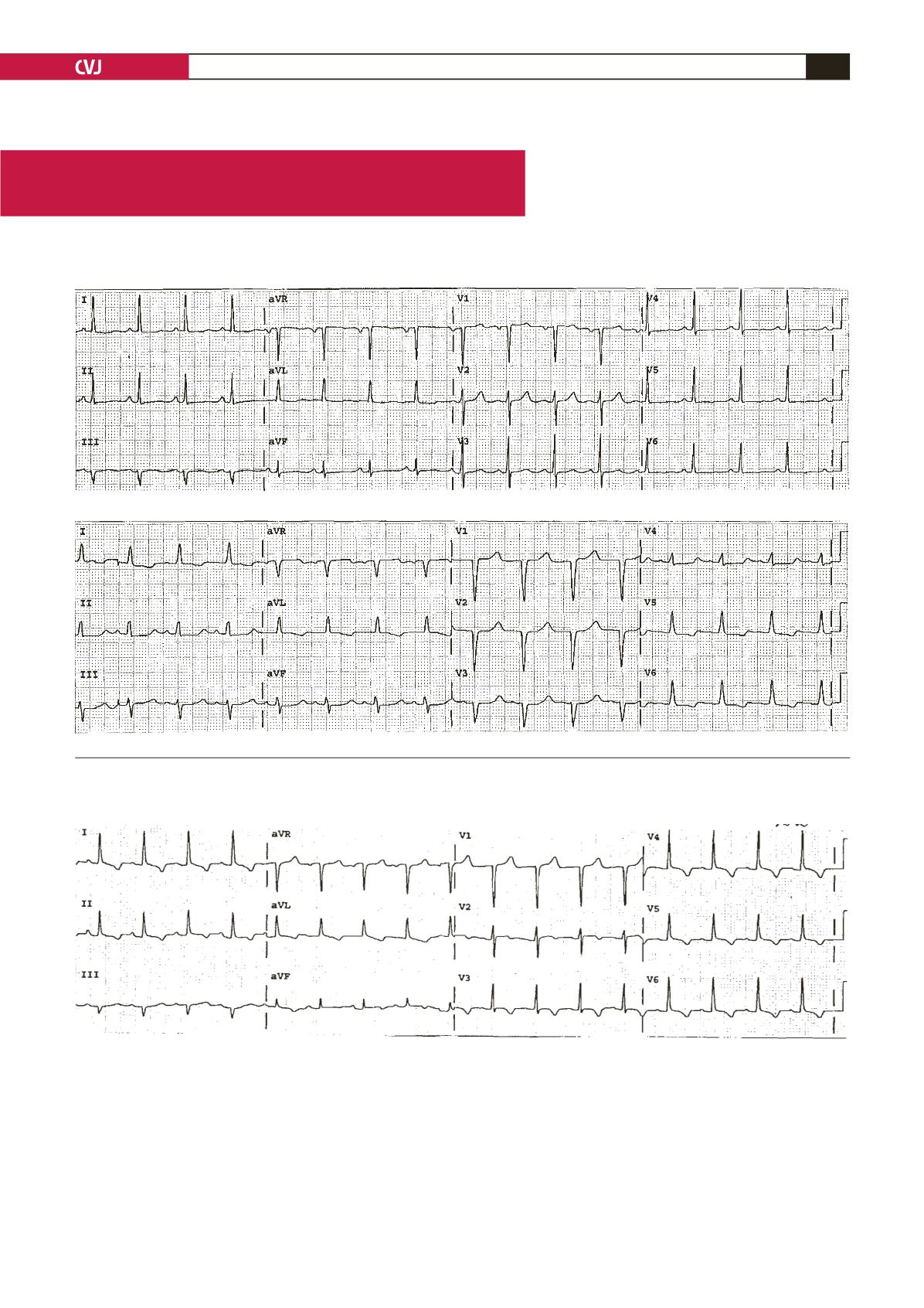
This is the ECG of a 54-year-old woman. What are your observations?
Is there a conduction abnormality or any evidence of MI?
Answer
What happened to the MI?
Here is an interesting sequence of tracings that may provide a modicum of humility! The top one is abnormal showing generalised
T-wave flattening and a long QT interval… ho-hum … The bottom one shows that the QRS duration has increased and the tiny Q
waves in leads I and AVL are no longer present – the pattern of incomplete LBBB. The QT remains prolonged and now the R waves
in VI3 have vanished… new anterior MI?
The tracing below is the next day. The incomplete LBBB has resolved and the R waves have returned, removing concern about
the anterior infarction.
Explanation: normally left-to-right septal activation results in an initial vector that is directed anteriorly and is dependent on the
integrity of the left bundle branch. In the presence of incomplete LBBB, this wave front can change direction and proceed from right
to left (anterior to posterior), removing the R waves that should be present in V1, 2, 3.
If you anticipated this possibility, you will be awarded a gold star …if not, stay alert.


