


 58 / 68

CARDIOVASCULAR JOURNAL OF AFRICA • Volume 26, No 1, January/February 2015
e8
AFRICA
Case Report
Treatment of an unusual complication of transfemoral
TAVI with a new technique: successful occlusion of
ventricular septal defect by opening the closure device
in the ascending aorta
Huseyin Dursun, Cenk Erdal, Oktay Ergene, Baris Unal, Zulkif Tanriverdi, Dayimi Kaya
Abstract
Ventricular septal defect (VSD) is a rare complication of
transcatheter aortic valve implantation (TAVI) via the trans-
femoral approach. Aetiological factors leading to VSD have
been reported as post-balloon dilatation, oversized prosthesis
implantation, and severe calcification of the aorta. However,
we present a case of VSD occurring after TAVI with an
Edwards Sapien XT prosthesis without any distinct aetiologi-
cal factors. We used a new technique for closure of the signifi-
cant VSD; opening the left ventricular disc of the closure
device in the ascending aorta and successfully implanting the
device without any damage to the bioprosthetic valve.
Keywords:
transcatheter aortic valve implantation, complication,
ventricular septal defect
Submitted 2/6/14, accepted 2/12/14
Published online 2/12/14
Cardiovasc J Afr
2015;
26
: e8–e10
www.cvja.co.zaDOI: 10.5830/CVJA-2014-077
Ventricular septal defect (VSD) is one of the rare complications
of transfemoral transcatheter aortic valve implantation (TAVI).
1-2
In the literature there are four reported cases using the Edwards
Sapien XT prosthesis (Edwards Lifesciences, Irving CA) and
one case with a CoreValve ReValving system (Medtronic, Irvine,
California).
The most prominent aetiological factors for VSD formation
are reported as post-balloon dilatation, oversized prosthesis
implantation, and severe calcification of the aorta.
3-7
However,
in this report, we present a VSD occurring after transfemoral
implantation of an Edwards Sapien XT prosthesis, without any
of these aetiological factors.
The VSD was successfully occluded retrogradely with a new
technique. To our knowledge this report is the first describing
successful closure of a VSD after TAVI with an Edwards Sapien
XT prosthesis. We used a unique technique for the closure
procedure.
Case report
A 73-year-old woman with dyspnoea in NYHA functional class
III was referred to our institution for severe aortic stenosis.
Transthoracic echocardiography (TTE) of the patient showed
a calcific aortic valve with an area of 0.7 cm
2
, and a mean
gradient of 45 mmHg. The aortic annulus diameter measured
by multislice computed tomography (CT) was 23 mm. There was
no critical stenosis on coronary and peripheral angiography with
a femoral artery diameter of 7 mm. Her logistic EuroSCORE
was 33.8%.
Department of Cardiology, Faculty of Medicine, Dokuz
Eylul University, Izmir, Turkey
Huseyin Dursun, MD,
drhuseyindursun@gmail.comOktay Ergene, MD
Baris Unal, MD
Zulkif Tanriverdi, MD
Dayimi Kaya, MD
Department of Cardiovascular Surgery, Faculty of
Medicine, Dokuz Eylul University, Izmir, Turkey
Cenk Erdal, MD
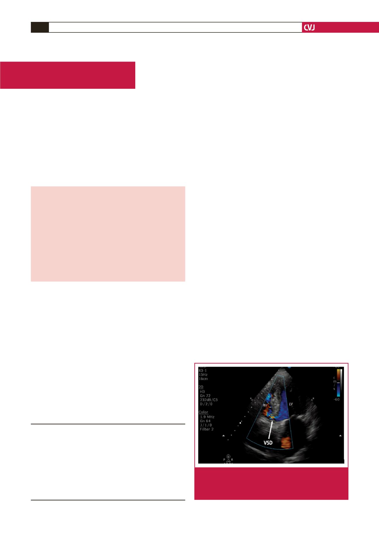
Fig. 1.
Transthoracic apical four-chamber view showing left-
to-right shunt at the interventricular septum level. VSD
= ventricular septal defect; RV = right ventricle, LV =
left ventricle.