
CARDIOVASCULAR JOURNAL OF AFRICA • Volume 28, No 1, January/February 2017
AFRICA
e3
been reported. The second interesting abnormality was the
PM. It has been suggested that isolated PM hypertrophy is
a possible variant of HCM, but only a few cases have been
reported in the literature.
3-6
Some of these patients presented with
electrocardiographic findings, such as high left precordial voltage
and inverted T waves, especially those with posteromedial
PM hypertrophy.
3,5
In our case, hypertrophy and malposition
occurred at the anterolateral PM.
Treatment of HCM is based on the anatomical abnormality.
In a subset of patients with HCM, LVOT obstruction will be
present not only because of septal hypertrophy, but also owing
to muscular apposition created by the abnormal PM. Failure to
recognise this anomaly would not relieve the obstruction.
7
In our case, aside from the abnormal PMand IVS, the chordae
tendineae connecting them with the mitral valve also contributed
to the crowded LVOT. Pre-operative identification of these three
contributors to LVOT obstruction altered the surgical strategy.
Therefore, this patient underwent septal myectomy of the sharp
angle, partial PM resection in the LVOT, as well as removal of
the abnormal chordae tendineae. Excessive PM resection could
have caused mitral valve insufficiency, which may have resulted
in the need for mitral valve replacement or surgical mitral leaflet
manipulation. Fortunately, the saline injection test showed only
trivial mitral valve regurgitation.
Redaelli
et al
.
8
proposed a procedure to reposition the
anterior PM followed by adjunctive implantation of a complete
semi-rigid mitral ring to abolish the systolic anterior motion and
residual mitral insufficiency. If our patient had had moderate to
severe mitral regurgitation, a semi-rigid mitral ring or even mitral
valve replacement would have been considered.
Conclusion
Different mechanisms causing LVOT obstruction may occur in
subgroups of patients with HCM. The mechanism of this rare
LVOT obstruction resulted from focal basal IVS hypertrophy and
angulation deformity, and abnormality of the PM and chordae
tendineae. Although we did not have genetic evidence, this
abnormal combination may represent a gap in our knowledge
of HCM. Careful echocardiographic and other radiological
assessment is needed before surgery, which could change the
diagnosis and management of HCM.
References
1.
Arad M, Seidman JG, Seidman CE. Phenotypic diversity in hypertroph-
ic cardiomyopathy.
Hum Mol Genet
2002;
11
(20): 2499–2506.
2.
Maron BJ, Sherrid MV, Haas TS, Lindberg J, Kitner C, Lesser JR.
Novel hypertrophic cardiomyopathy phenotype: segmental hypertrophy
isolated to the posterobasal left ventricular free wall.
Am J Cardiol
2010;
106
(5): 750–752.
3.
Kobashi A, Suwa M, Ito T, Otake Y, Hirota Y, Kawamura K. Solitary
papillary muscle hypertrophy as a possible form of hypertrophic cardio-
myopathy.
Jpn Circ J
1998;
62
(11): 811–816.
4.
Ta
ş
demir O, Küçükaksu DS, Kural T, Bayazit K. Hypertrophic obstruc-
tive cardiomyopathy in combination with anomalous insertion of papil-
lary muscle directly into anterior mitral leaflet and ‘sawfish’ systolic
narrowing of the left anterior descending coronary artery.
Tex Heart
Inst J
1994;
21
(4): 317–320.
5.
Ferreira C, Delgado C, Vázquez M, Trinidad C, Vilar M. Isolated
papillary muscle hypertrophy: A gap in our knowledge of hypertrophic
cardiomyopathy.
Rev Port Cardiol
2014;
33
(6): 379.e1–5.
6.
Correia AS, Pinho T, Madureira AJ, Araujo V, Maciel MJ. Isolated
papillary muscle hypertrophy: a variant of hypertrophic cardiomyo-
pathy? Do not miss a hypertrophic cardiomyopathy.
Eur Heart J
Cardiovasc Imaging
2013;
14
(3): 296.
7.
Maron BJ, Nishimura RA, Danielson GK. Pitfalls in clinical recognition
and a novel operative approach for hypertrophic cardiomyopathy with
severe outflow tract obstruction due to anomalous papillary muscle.
Circulation
1998;
98
(23): 2505–2508.
8.
Redaelli M, Poloni CL, Bichi S, Esposito G. Modified surgical approach
to symptomatic hypertrophic cardiomyopathy with abnormal papillary
muscle morphology: Septal myectomy plus papillary muscle reposition-
ing.
J Thorac Cardiovasc Surg
2014;
147
(5): 1709–1711.
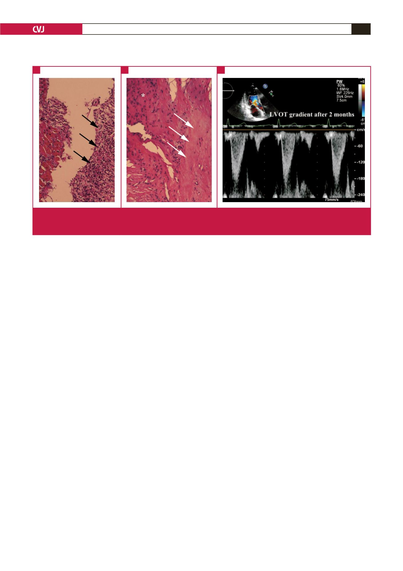
Fig. 3.
Histological examination showed inflammation (A, black arrows), cardiomyocyte hypertrophy and disarray (B, *), as well as
interstitial fibrosis (B, white arrows) (haematoxylin and eosin, ×100). At the two-month follow up, transthoracic echocardiog-
raphy demonstrated a LVOT gradient of 23 mmHg (C).
A
B
C


