


 72 / 76

CARDIOVASCULAR JOURNAL OF AFRICA • Volume 29, No 3, May/June 2018
e2
AFRICA
perform a routine myocardial biopsy, which was necessary for
the pathological diagnosis of myocarditis.
We had no choice but to send the patient back to the intensive
care unit for stabilisation of the haemodynamic status. However,
his level of consciousness did not improve (Glasgow coma
scale score 3). Emergency brain computed tomography (CT)
showed no intracranial haemorrhage, and magnetic resonance
imaging (MRI) was contraindicated because infusion pumps
were implanted. We were therefore unable to rule out acute stroke.
Meanwhile, 24-hour hypothermia therapy (HT) at
approximately 34°C was employed for neurological protection
in post-resuscitation circulatory shock. Transthoracic
echocardiogram showed general hypokinesia of both ventricles
(left ventricular ejection fraction 15–20%). Despite these
interventions, acute pulmonary oedema and deteriorating liver
and renal function with progressive oliguria ensued.
To avoid multiple organ dysfunction syndrome, a continuous-
flow Levitronix
®
CentriMag Bi-VAD (Levitronix
®
, Waltham,
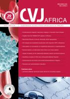
MA) was implanted via a median sternotomy under the guidance
of transoesophageal echocardiography. First, the left heart vent
tube was inserted from the right superior pulmonary vein into
the left ventricular apex, and the arterial cannula was inserted
into the ascending aorta. Second, the right heart vent tube
was inserted into the right atrium, and the arterial cannula
was inserted into the pulmonary artery (Fig. 1). Then purse-
string sutures with non-absorbable retention sutures secured
with tourniquets and spigots were tied around all the cannulae.
The vital signs immediately stabilised with Bi-VAD support
and the high-dose inotropic support was tapered off the next
day (dopamine: 5 mcg/kg/min, dobutamine: 5 mcg/kg/min and
norepinephrine: 3.7 mcg/min).
Because a Bi-VAD was inserted, systemic heparinisation
therapy was administered through a peripheral line to maintain an
activated clotting time (ACT) at 160–180 s using the Hemochron
®
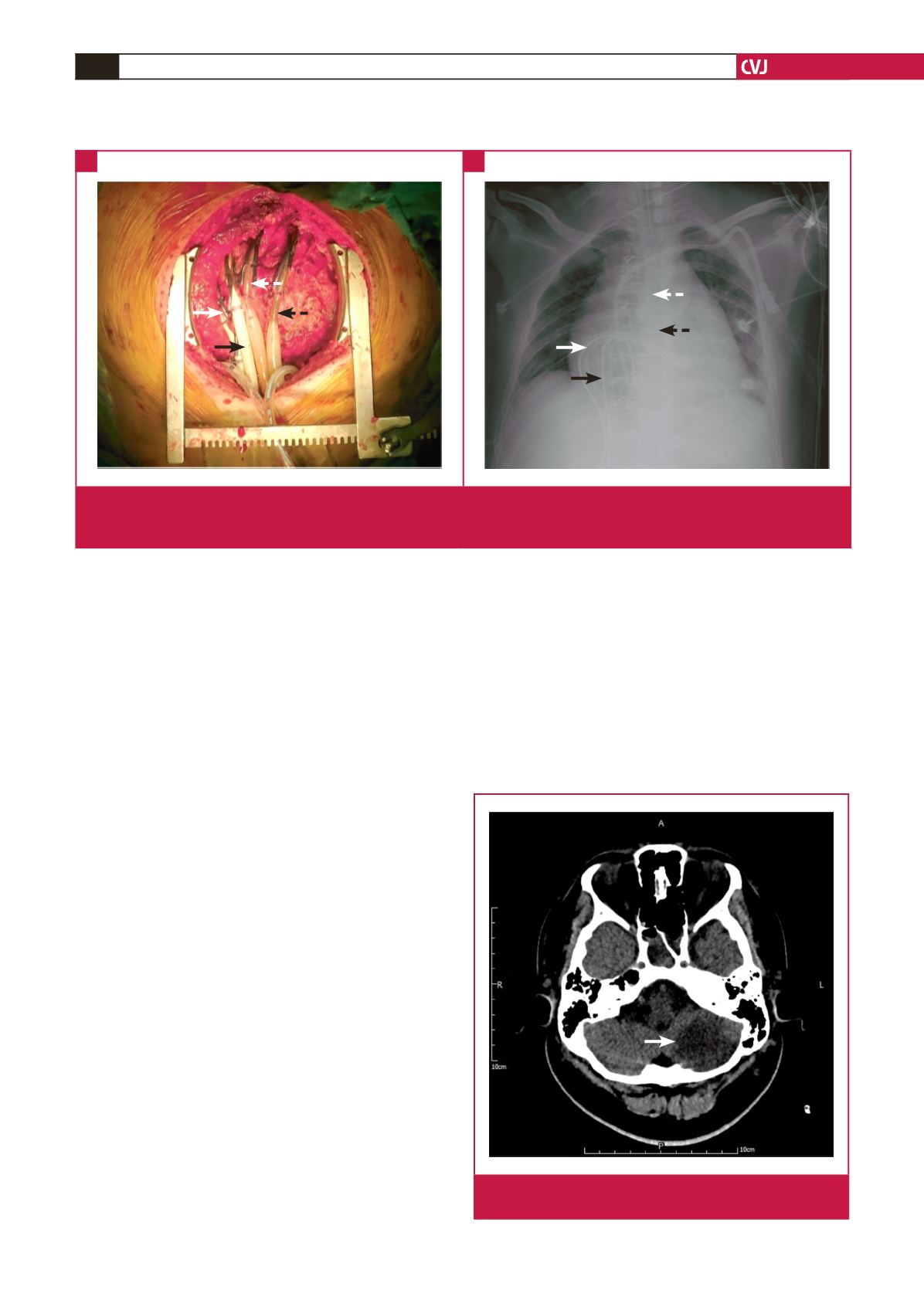
Response ACT point-of-care testing system. Unfortunately, CT the
following day showed an acute left cerebellar infarction (Fig. 2),
which resulted in right hemiplegia. We assumed that the cerebellar
infarction was caused by the cardiopulmonary resuscitation rather
than VAD-related thrombus formation. To prevent post-infarct
haemorrhage, we tapered the ACT to 140–160 s.
Three days later, the patient fully recovered from the coma,
and his muscle power also improved. Daily echocardiography
examinations showed progressive improvement of the cardiac
systolic function.
When the left ventricular ejection fraction was approximately
50%, the Bi-VAD was weaned (right-VAD: 0.5 l/min; left-VAD:
0.8 l/min). The patient underwent successful Bi-VAD removal,
Fig. 1.
(A) Photograph of the cardiac operation and (B) a chest X-ray showing the left heart venous drainage tube (white arrow),
the arterial perfusion tube (white dotted arrow), the right heart venous drainage tube (black arrow), and the arterial perfusion
tube (black dotted arrow).
A
B
Fig. 2.
Hypodense defect indicates acute left cerebellar
infarction (white arrow).