


 16 / 74

CARDIOVASCULAR JOURNAL OF AFRICA • Volume 30, No 2, March/April 2019
82
AFRICA
potential of –20 mV, the I
Na,Late
densities of the three mutations
increased from 0.81
±
0.03 pA/pF for the WT to 5.02
±
0.13 pA/
pF for F1473S, 594
±
0.47 pA/pF for T535I, and 4.12
±
0.12 pA/
pF for E1784K, respectively (
p
<
0.01,
n =
10). After exposure to
30
μ
M All, the I
Na,Late
densities of SCN5A mutations decreased
to 1.08
±
0.02 pA/pF for F1473S, 1.32
±
0.50 pA/pF for T535I,
and 0.97
±
0.31 pA/pF for E1784K, respectively (
p
<
0.01,
n =
10; Fig. 6B).
The effects of different concentrations of All on I
Na,Late
of the
SCN5A mutations were investigated. I
Na,Late
was inhibited by All
in a concentration-dependent manner. IC
50
was 15.2
±
2.2
μ
M
for F1473S, 41.8
±
3.6
μ
M for T535I, and 18.1
±
3.2
μ
M for
E1784K, respectively. The Hill coefficients were 0.87, 1.29 and
0.98, respectively (Fig. 6C).
Discussion
In epidemiological studies, the risk of AF was estimated to
be 1.42 times higher in hypertensive patients compared with
normotensives. Hypertension, age and diabetes are prominent risk
factors for AF.
11,12
Normally, the sodium channel was inactivated
within a few milliseconds of depolarisation. Some channels
remained open, creating a small but persistent influx of Na
+
throughout the plateau of AP during pathological conditions.
13,14
Especially in chronic physiological and pathological processes
(such as in the case of aging, myocardial hypertrophy, sick
sinus syndrome and heart failure), sodium channels may be
remodelling. Chang
et al
. found down-regulation of Nav1.5
protein expression and reduced I
Na
density in failing hearts
and ischaemia–reperfusion injury. Nav1.5 contributes to
arrhythmogenesis in heart failure due to the generation of I
Na,Late
.
15
Up-regulated Nav1.8 augmented I
Na,Late
in human hypertrophied
myocardium and prolonged the APD.
16,17
Sick sinus syndrome is a common arrhythmia often associated
with aging or organic heart diseases. The disease-causing gene
is closely related to the sodium channel.
18
The SHR cells
showed electrophysiological remodelling of the left atrium,
leading to increased vulnerability to burst pacing-induced atrial
arrhythmias.
2
Our investigation demonstrated larger I
Na,Late
and longer APD
in SHR atrial cells compared with WKY cells. The magnitude
of I
Na,Late
may increase significantly in chronic pathological
settings. Sossalla
et al
.
19
reported that I
Na,Late
increased in atrial
myocytes isolated from the right atrial appendage of persistent
AF patients. I
Na,Late
densities in left atria have also been reported
to increase in a rabbit left ventricular hypertrophy model caused
by hypertension.
13
I
Na,Late
contributes to the plateau phase of the cardiac AP and
is related to arrhythmogenesis under pathological conditions.
Although the contribution is small relative to the peak current,
I
Na,Late
cannot be neglected. A small, persistent Na
+
current
prolongs the plateau APD and induces a Na
+
load that may
indirectly increase intracellular Ca
2+
concentrations. Both AP
prolongation and Ca
2+
overload are reported to be the main
causes of AF.
20,21
Our findings suggest that enhanced I
Na,Late
is
involved in the occurrence and development of AF.
We also found that the window currents of SHR atrial cells
were enhanced. Several factors contribute to the late sodium
WKY
SHR
All 30
μ
M
WKY
SHR All 30
μ
M
I
Na,late
/I
peak
(%)
1.0
0.8
0.6
0.4
0.2
0.0
**
##
WKY
SHR All 30
μ
M
I
Na,peak
(pA/pF)
0
–50
–100
–150
–200
–250
Log [C]
μ
M
0.1
1
10
100 1000
Fraction of maximum
1.0
0.8
0.6
0.4
0.2
0.0
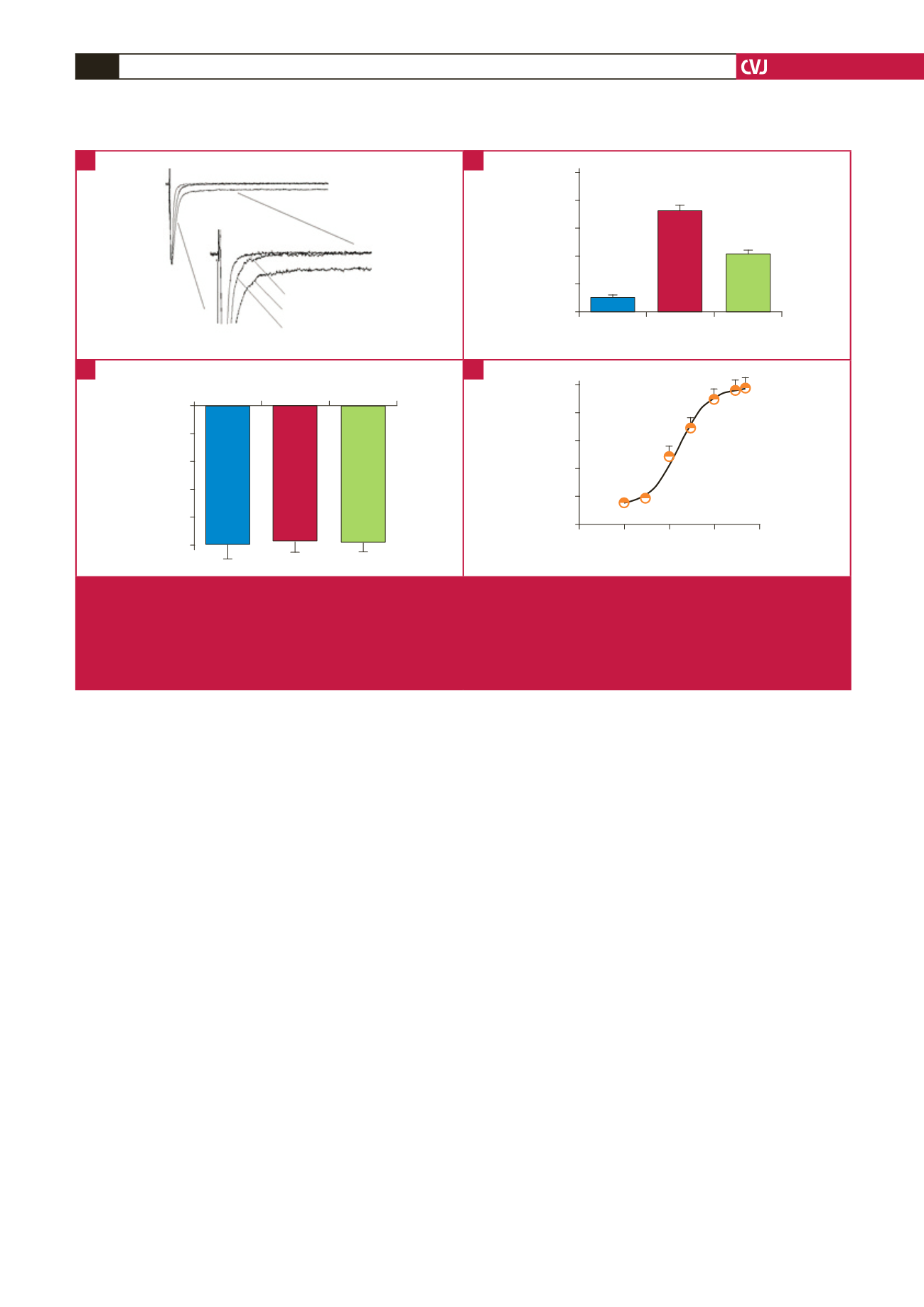
Fig. 3.
Effect of All on late sodium current (I
Na,Late
) of SHR myocytes. (A) Representative current traces recorded from WKY and
SHR cells with 30
μ
M All. I
Na,Late
of SHR myocytes was significantly larger than that of WKY cells, which was inhibited by
All. (B) Incremental ratio of I
Na,Late
/I
Na,peak
in SHR cells reduced from 0.71
±
0.02 to 0.37
±
0.02% and ended at 0.09
±
0.01%
of WKY cells. (C) At a test potential of –20 mV, I
Na,peak
did not change in the three groups. (D) I
Na,Late
was inhibited by All in a
concentration-dependent manner. IC
50
was 16.8
±
2.2
μ
M, Hill coefficient: 0.96 (
n
= 15). **
p
<
0.01 vs WKY group.
##
p
<
0.01
vs SHR group.
A
C
B
D