CARDIOVASCULAR JOURNAL OF AFRICA • Vol 24, No 5, June 2013
172
AFRICA
examined monthly as out-patients. During these follow-up visits,
we were watching for fever, dyspnoea, and signs of right heart
failure or inter-current illness.
Data analysis was done with SPSS. The results are expressed
as numbers and percentage.
Results
During the study period, 48 patients with infective endocarditis
were admitted to hospital, including 14 (29.1%) with right-sided
endocarditis. The mean age was 25.5
±
12.5 years (range from
9–80 years) and the gender ratio of women to men was 2.3.
Children accounted for half of the right-sided endocarditis (seven
cases).
A peripheral venous access had been performed in 12 patients
in primary healthcare facilities and nine others had received
inadequate antibiotic treatment. No case of drug addiction was
recorded. Three patients were HIV positive while four were in
the post-partum period.
Venous access was the entry point for bacteria in 12 patients
(85.7%). The indications of venous access were malnutrition
(five cases), childbirth (four cases), sickle cell crisis (two cases)
and malaria (one case). In the other two cases, no entry point
was found.
The clinical features included infectious syndrome in all
patients, and right heart failure in nine cases. Tricuspid syndrome,
consisting of fever associated with long-term lung damage
(usually asymptomatic), anaemia and microscopic haematuria
was found in six patients (42.8%). The diagnosis of infective
endocarditis was based on the association of two major criteria
in 12 patients and the association of one major criterion and three
minor criteria in two other cases.
Blood sample cultures were positive in 11 patients, isolating
Streptococcus pneumonia
in six cases,
Staphylococcus aureus
in
three cases and
Hemophilus influenzae
in two cases. Anaemia
was common, as well as biological inflammatory syndrome
(raised CRP, hyper-fibrinaemia and accelerated sedimentation
rate) and leucocytosis.
The electrocardiogram revealed a left atrial enlargement
in seven patients, left ventricular hypertrophy in six patients,
right atrial and ventricular hypertrophy in three cases and atrial
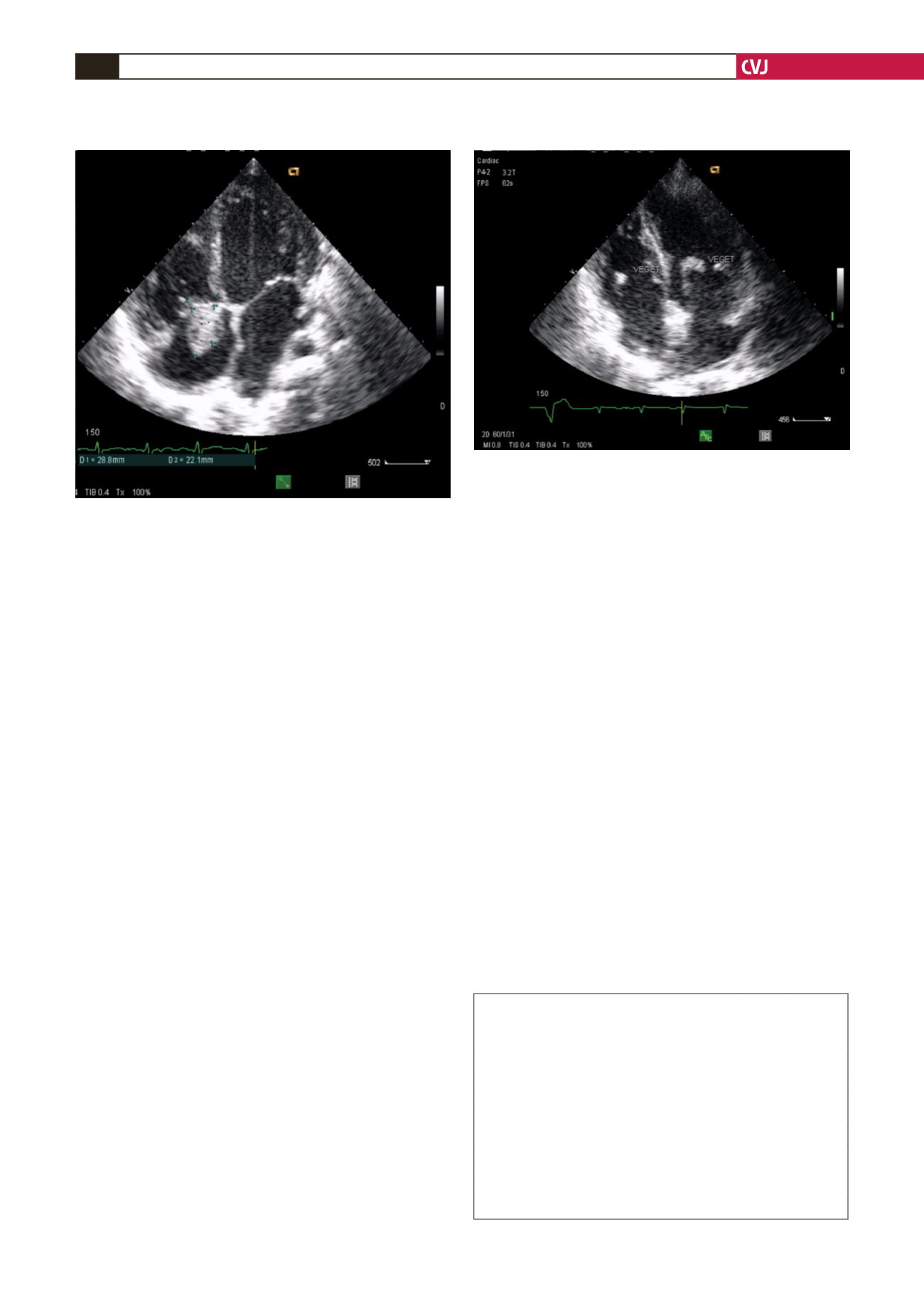
fibrillation in two children. Doppler echocardiography revealed
vegetations in all patients. Vegetations were localised in the
right heart only in 11 cases and on the tricuspid valve only in
seven cases (Fig. 1). Otherwise vegetations were found both on
the tricuspid and mitral valves in two cases (Fig. 2) and on the
pulmonary valves in one case (Fig. 3). The average surface area
of the vegetations was 2.9
±
0.6 cm
2
(range 1.2–5.2). All patients
had both tricuspid and pulmonary regurgitation.
Underlying heart diseases diagnosed by echocardiography
Doppler are listed in Table 1.
Prior to the results of blood sample cultures, an early treatment
with probabilistic antibiotics was made of a combination of a
third-generation cephalosporin and an aminoglycoside, except in
one case where the aminoglycoside was not introduced because
of kidney failure. Once blood samples cultures had revealed a
pathogen, the antibiotic treatment was then adjusted according
to the antibiogram. Treatment was therefore adjusted in five
patients. Heart failure was treated as appropriate.
The average hospital stay was 35
±
7 days (range 24–49 days).
The clinical course was marked by a lowering of temperature
within an average treatment period of 10 days. Heart failure
symptoms decreased as well. One fatality was reported in a child
after 14 days of hospitalisation due to septic shock. After a mean
TABLE 1. DISTRIBUTION OF UNDERLYING HEART DISEASE IN
PATIENTSWITH RIGHT-SIDED HEART ENDOCARDITISAT
THEYALGADO OUEDRAOGO UNIVERSITY HOSPITAL
FROM JANUARY 2010 TO DECEMBER 2011 (
n
=
14)
Underlying heart disease
Number Percentage
Peripartal cardiomyopathy
4
28.6
Dilated cardiomyopathy
2
14.3
Ventricular septal defect
2
14.3
Pulmonary stenosis
+
inter-atrial communication
2
14.3
Tetralogy of Fallot
1
7.1
Restrictive ventricular septal defect
+
ductus arteriosus
1
7.1
No heart disease found
2
14.3
Total
14
100
Fig. 1. Two-dimensional four-chamber echocardiogram
showing large vegetations on the tricuspid valve.
Fig. 2. Two-dimensional four-chamber echocardiogram
showing vegetations on both tricuspid and mitral valves.


