


 55 / 64

CARDIOVASCULAR JOURNAL OF AFRICA • Volume 31, No 5, September/October 2020
AFRICA
275
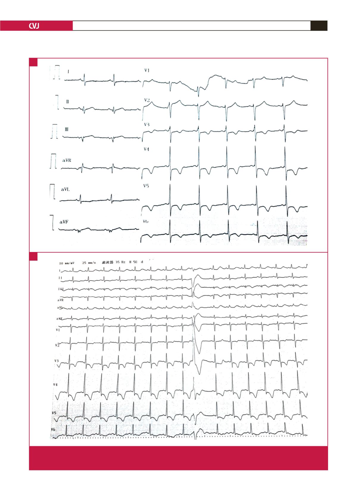
Fig. 1.
Twelve-lead ECG recordings. A: ECG on the first admission shows an obvious ST-segment depression with giant inverted
T waves in leads V
3
–V
6.
B: ECG on the second admission shows an obvious ST depression and inverted T waves in leads
V
1
–V
6
, associated with a premature ventricular contraction.
A
B