


 61 / 64

CARDIOVASCULAR JOURNAL OF AFRICA • Volume 31, No 5, September/October 2020
AFRICA
281
Internal thoracic artery pseudoaneurysm after redo
aortic root replacement
Yoshinori Kuroda, Tetsuro Uchida, Azumi Hamasaki, Mitsuaki Sadahiro
Abstract
Pseudoaneurysm of the internal thoracic artery (ITA) or
bleeding from the ITA is an extremely rare complication
after cardiovascular surgery via a median sternotomy. Early
treatment is needed in the case of massive haemorrhage or a
rapidly enlarging pseudoaneurysm. Herein, we present a rare
case of a delayed large pseudoaneurysm of the right ITA in a
49-year-old woman with Marfan syndrome who underwent
redo aortic root replacement via re-median sternotomy and
pacemaker implantation. Diagnostic selective angiography
revealed the origin of the pseudoaneurysm, and simultane-
ous transcatheter embolisation of the ITA was successfully
performed. Follow-up computed tomography imaging showed
no evidence of contrast media extravasation from the ITA and
recurrent extra-pleural haemorrhage. Our findings suggest
that postoperative management of patients who have under-
gone median sternotomy, including cardiovascular surgeries,
should also focus on the prevention or early detection of pseu-
doaneurysm of the ITA to avoid life-threatening conditions.
Keywords:
Marfan syndrome, post aortic root replacement, pseu-
doaneurysm of the internal thoracic artery
Submitted 24/9/19, accepted 28/5/20
Published online 16/6/20
Cardiovasc J Afr
2020;
31
: 281–282
www.cvja.co.zaDOI: 10.5830/CVJA-2020-014
Pseudoaneurysm or bleeding of the internal thoracic artery (ITA)
is an extremely rare complication after cardiovascular surgery via
a median sternotomy, especially during the delayed postoperative
phase. Early treatment may be needed in the case of massive
haemorrhage or a rapidly enlarging pseudoaneurysm. Herein, we
describe a case of pseudoaneurysm of the ITA in a patient with
Marfan syndrome after redo aortic root replacement.
Case report
The patient was a 49-year-old woman who underwent valve-
sparing aortic root replacement (re-implantation procedure)
and total arch replacement for a type A aortic dissection.
Although she was asymptomatic, four months after the aortic
root replacement, a massive aortic root pseudoaneurysm was
detected on routine follow-up computed tomography (CT)
scan. Redo aortic root replacement (Bentall procedure) through
re-median sternotomy was urgently performed.
After the surgery, complete atrioventricular block occurred,
and pacemaker implantation (PMI) was performed via the
right axillary vein through a cut-down procedure two days
after the redo aortic root replacement. On the following day,
chest radiography revealed massive right pleural effusion (Fig.
1A) although chest radiography performed the day before PMI
revealed little pleural effusion (Fig. 2). A large amount of blood
(1 600 ml) was drained by thoracentesis.
Emergency CT conducted immediately after thoracentesis
revealed upper anterior extra-pleural haemorrhage, extravasation
of contrast media inside the haemorrhage and pleural effusion
(Fig. 1B). Although the extravasation inside the extra-pleural
haemorrhage detected on CT looked like a torturous abnormal
vessel, pre-operative CT revealed a normal ITA and its branch
vessels (Fig. 3). Additionally, the origin of the extravasation was
unclear on CT. Selective angiography of the right ITA detected
extravasation from a thin branch of the right ITA running to the
mediastinum (Fig. 4A). Right ITA embolisation was completed
with coils and n-butyl-2-cyanoacrylate, and the bleeding was
controlled (Fig. 4B).
The patient had an uneventful postoperative recovery, and
follow-upCT showed no evidence of contrast media extravasation
from the ITA or recurrent extra-pleural haemorrhage.
Discussion
Injury and pseudoaneurysm or bleeding of the ITA are
reported as rare postoperative complications of surgery via a
median sternotomy, which are caused by closure wire injury,
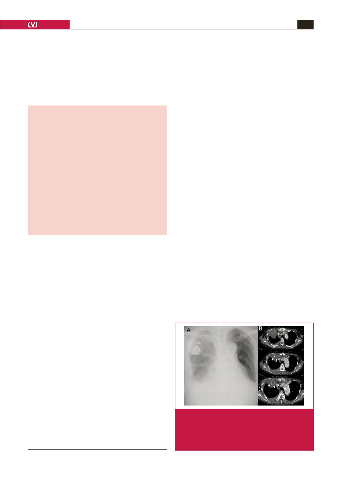
Fig. 1.
Chest radiography and computed tomography after
pacemaker implantation. A: Chest radiograph shows
massive right pleural effusion. B: Emergency computed
tomography after thoracentesis shows extravasation of
contrast to the extra-pleural haemorrhage of the upper
portion of the right pleura (arrow), and pleural effusion.
Division of Cardiovascular Surgery, Department of Surgery
II, Faculty of Medicine, Yamagata University, Yamagata, Japan
Yoshinori Kuroda, PhD,
y-kuroda@med.id.yamagata-u.ac.jpTetsuro Uchida, PhD
Azumi Hamasaki, PhD
Mitsuaki Sadahiro, PhD