


 31 / 60

CARDIOVASCULAR JOURNAL OF AFRICA • Volume 32, No 2, March/April 2021
AFRICA
85
mechanism with SARS-COV because the two viruses are highly
homologous in the genome.
13,14
In our study, plasma TnT levels were significantly positively
correlated with other plasma inflammatory markers such as
fibrinogen, D-dimer, ferritin, procalcitonin and CRP, indicating
that myocardial injury may be closely related to inflammatory
pathogenesis during the evolution of the disease.
Viral particles could precipitate a cytokine storm and a
series of immune responses. Huang
et al
.
4
emphasised that
in patients with COVID-19, an imbalance of T-helper 1 and
T-helper 2 responses developed in a cytokine storm, which
may have contributed to myocardial injury. The release of
inflammatory cytokines after infection may cause a reduction in
coronary blood flow, decrease in oxygen supply, destabilisation
of coronary plaque and microcirculatory thrombogenesis.
Unpredictably, the proportion of smokers was statistically
significantly lower in patients with myocardial injury than in
those without injury (
p
<
0.01) (Table 1).
In a recently published article, Guo
et al
.
15
found 11 of 18
patients who were in the smoking group had normal TnT levels
and seven had elevated troponin levels. However, it was not
statistically significant. In another trial, researchers found no link
between cigarette smoking and the severity of COVID-19 among
cases in China, according to results of a preliminary meta-
analysis published in the
European Journal of Internal Medicine
.
16
Although there are articles showing no relationship between
smoking and the severity of COVID-19, there are also articles
showing the opposite. Therefore, this situation remains uncertain
and more studies are needed to clarify it.
Study limitations
First, as a retrospective study, information regarding cardiovascular
complications, such as from echocardiography, and other
inflammatory markers, such as cytokine levels, including IL-6, are
not presented in this study because the data were incomplete owing
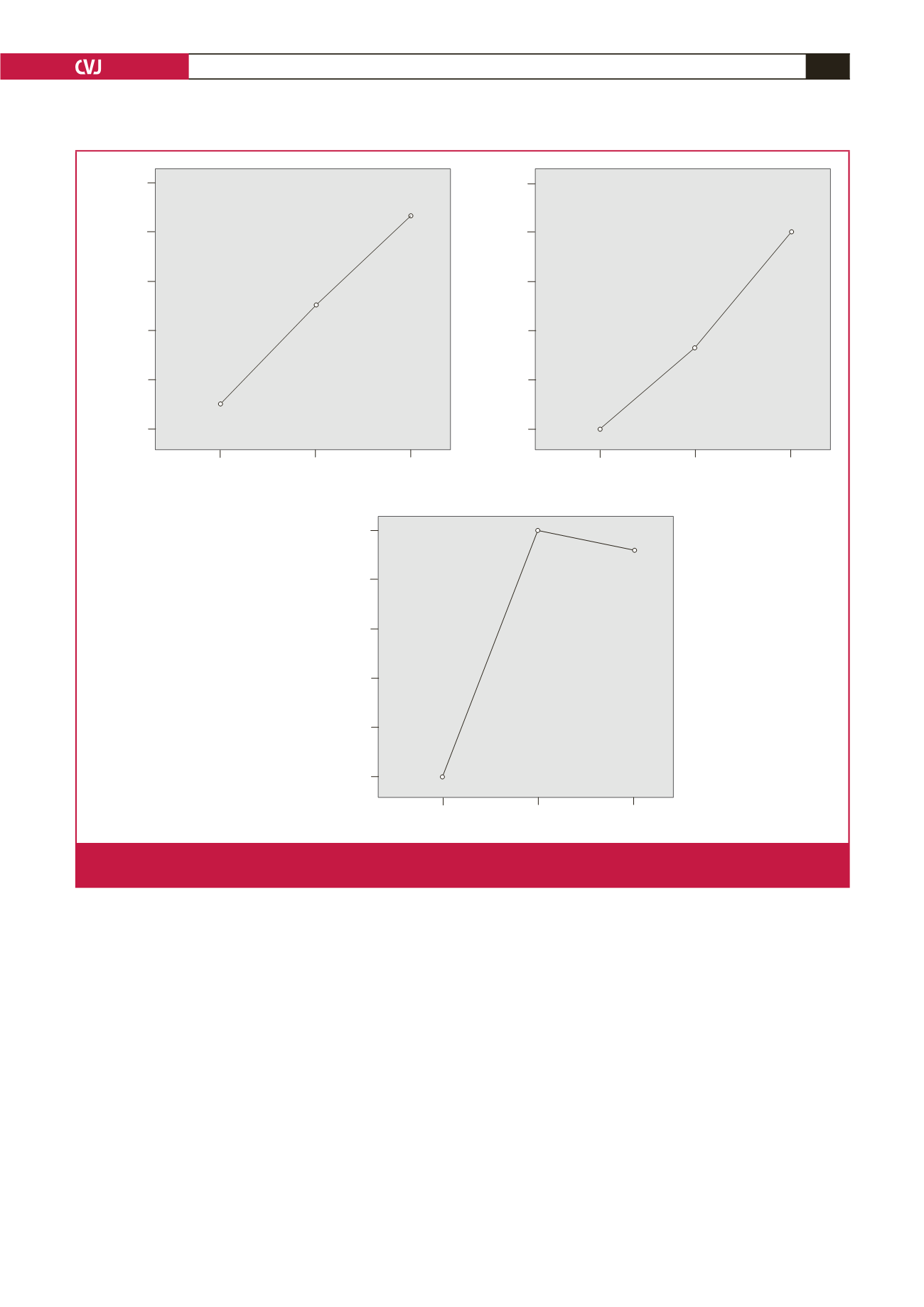
2 000.00
1 800.00
1 600.50
1 400.00
1 200.50
1 000.00
1.00 2.00 3.00
Thorax CT
Mean of D-dimer,
μ
g/l
Thorax CT: 1 (mild pneumonia), 2 (moderate pneumonia), 3 (severe pneumonia)
900.00
800.00
700.00
600.00
500.00
400.00
1.00 2.00 3.00
Thorax CT
Mean of ferritin, ng/ml
600.00
500.00
560.00
540.00
520.00
500.00
1.00 2.00 3.00
Thorax CT
Mean of fibrinogen, mg/dl
Fig. 4.
Mean plots of serum levels of ferritin, D-dimer and fibrinogen compared with patients grouped according to thorax CT scans,
which were divided into three categories: 1, mild pneumonia; 2, moderate pneumonia; and 3, severe pneumonia.