CARDIOVASCULAR JOURNAL OF AFRICA • Vol 21, No 4, July/August 2010
222
AFRICA
calcification coincided with the typical location of left ventricu-
lar aneurysms, which are usually located at the apex and often
involve the anterior and lateral walls. Trauma, cardioversion,
infection and endocardial fibrosis are rare causes of coarse,
amorphous myocardial calcifications, which are distinct from the
fine, curvilinear calcifications of a left ventricular aneurysm.
2
Similar previous case reports also demonstrated a calci-
fied LVA using left ventriculography, chest X-ray, cardiac
magnetic resonance imaging and computerised tomography.
3-5
Interestingly, however, we showed a calcified LVA appearing like
a huge halo image during coronary angiography.
References
1. Cabin HS, Roberts WC. True left ventricular aneurysm and healed
myocardial infarction.
Am J Cardiol
1980;
46
: 754–763.
2. Kazamias TM, Caine TH, Rowe GG, Crumpton CW. Calcification of the
interventricular septum.
J Am Med Assoc
1970;
213
: 1896–1898.
3. Charokopos N, Antonitsis P, Rouska E, Toumbouras M. Calcified aneu-
rysm of the left ventricle mimicking hydatid disease of the lung.
Eur J
Cardiothorac Surg
2008;
33
(5): 925.
4. Karaahmet T, Tigen K, Gurel E, Tanalp AC, Basaran Y.
Dystrophic
calcification of the aneurysmatic left ventricular apex.
Congest Heart
Fail
2009;
15
(4): 196–198.
5. Nakajima O, Sano I, Akioka H
.
Images in cardiovascular medicine.
Marked calcified left ventricular aneurysm.
Circulation
1997;
95
(7):
1974.
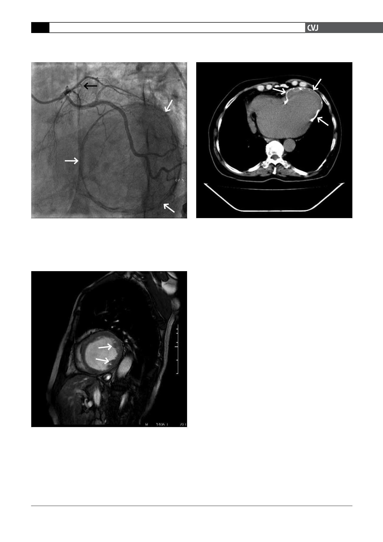
Fig. 3. Cardiac magnetic resonance imaging confirming
the left ventricle aneurysm with intramural thrombus
formation (the arrows show the thrombus formation in
left ventricle cavity).
Fig. 4. Cardiac computed tomography confirming the
left ventricle aneurysm (the arrows show the edge of the
calcified left ventricle aneurysm).
Fig. 2. Left anterior oblique coronary angiographic image
with caudal angulation showing total occlusion of the left
anterior descending artery and a halo in the heart after
left coronary artery injection. (The black arrow shows
the point of the left anterior descending artery occlusion
and the white arrows show the edge of the calcified left
ventricle aneurysm.)


