CARDIOVASCULAR JOURNAL OF AFRICA • Vol 22, No 5, September/October 2011
272
AFRICA
Isolated tricuspid valve prolapse: identification using
two- and three-dimensional echocardiography and
transoesophageal echocardiography
GONENC KOCABAY, DICLE SIRMA, MERAL MERT, KURSAT TIGEN
Abstract
We present a case of isolated prolapse of the tricuspid ante-
rior leaflet in an asymptomatic 34-year-old man who was
referred to our hospital for a routine check up.We performed
two-and three-dimensional transoesophageal echocardiog-
raphy (TEE). We found three-dimensional TEE a useful,
non-invasive tool that can provide additional information
to two-dimensional echocardiography in the assessment of
tricuspid valve prolapse.
Keywords:
isolated tricuspid valve prolapse
Submitted 17/12/10, accepted 16/2/11
Published online 27/6/11
Cardiovasc J Afr
2011;
22
: 272–273
DOI: 10.5830/CVJA-2011-006
Tricuspid valve prolapse is frequently found together with mitral
valve prolapse, and rarely as an isolated occurrence.
1,2
Isolated
prolapse of the valvular leaflets may cause significant tricuspid
regurgitation.
3
We present a case of isolated prolapse of the
tricuspid anterior leaflet in an asymptomatic 34-year-old man
who was referred to our hospital for a routine check-up. He
denied any blunt chest trauma such as a traffic accident.
Case report
On examination, there was a thrill and 4/6 pansystolic murmur in
the tricuspid area. His blood pressure was 120/70 mmHg and the
heart rate was regular and 90 beats per min. Electrocardiography
showed sinus rhythm with right bundle branch block morphol-
ogy. A 24-hour rhythm Holter examination was unremarkable.
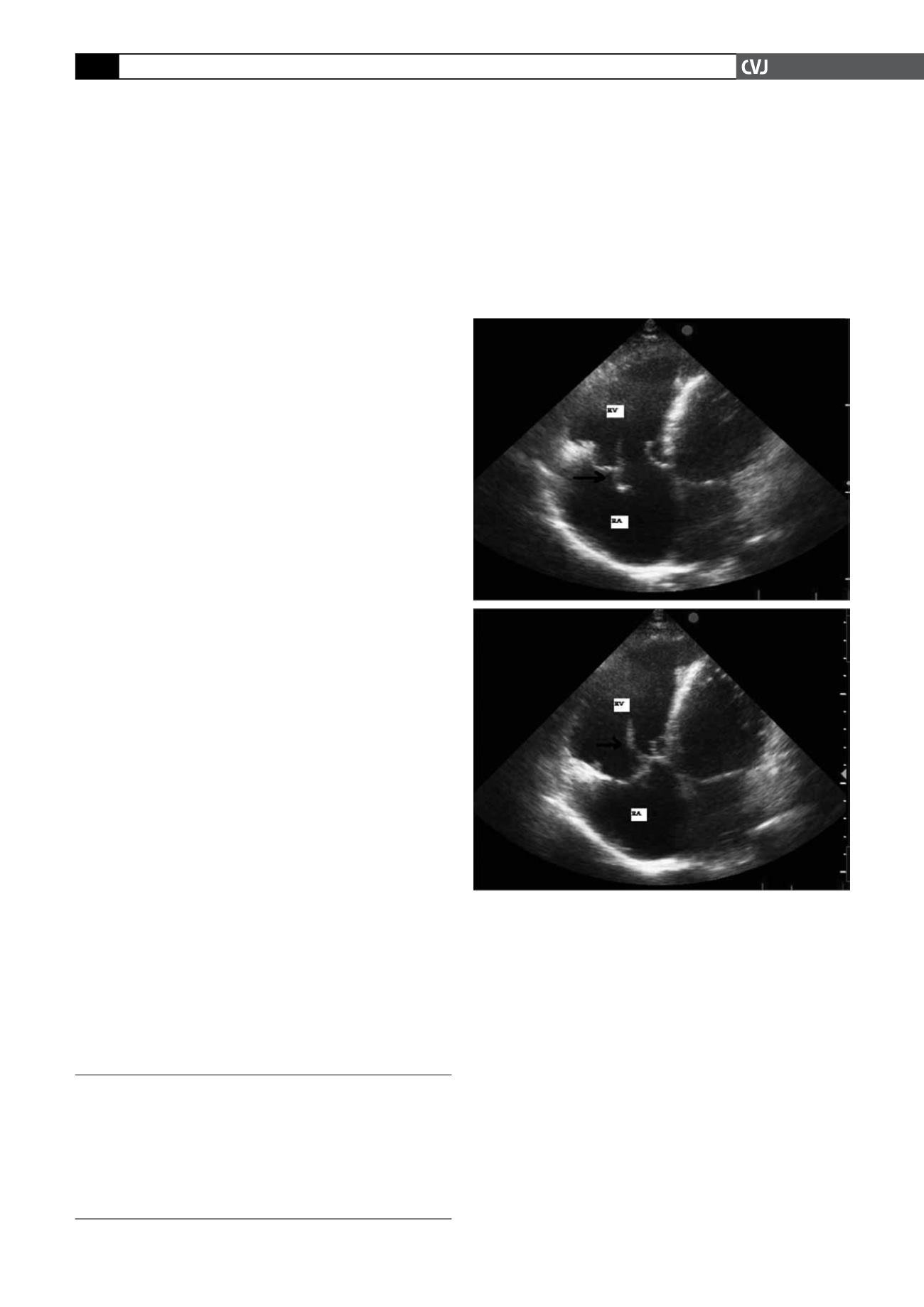
Echocardiographic evaluation showed an isolated prolapse
of the tricuspid anterior leaflet with severe tricuspid regurgita-
tion and right-sided heart chamber enlargement (Fig. 1). The
left heart chamber sizes and systolic function were normal.
Transoesophageal echocardiography (TEE) was performed to
better define the tricuspid valve structure. TEE also revealed
isolated anterior tricuspid valve prolapse with severe tricuspid
regurgitation and patent foramen ovale (PFO) with atrial septal
aneurysm (Fig. 2). The other valves were structurally and func-
tionally normal. We also performed three-dimensional TEE
(Fig. 3).
Due to the existence of a PFO and severe tricuspid regurgita-
tion, surgery was suggested. The tricuspid annulus was repaired
using a Carpentier-Edwards ring. A tissue patch was used to
repair the PFO.
Discusssion
Tricuspid valve prolapse is commonly associated with mitral
valve prolapse and is rarely an isolated occurrence. Isolated
severe tricuspid regurgitation can occur from isolated prolapse
of the valvular leaflets.
1
Two-dimensional echocardiography
using multiple views is an appropriate technique for the demon-
stration of tricuspid valve prolapse. The posterior leaflet is seen
only on the long-axis parasternal view.
4
As obtaining this view
Kartal Kosuyolu Heart and Research Hospital, Department
of Cardiology, Kartal, Istanbul, Turkey
GONENC KOCABAY, MD,
DICLE SIRMA, MD
KURSAT TIGEN, MD
Kayseri Education and Research Hospital, Department of
Endocrinology and Metabolism, Kayseri, Turkey
MERAL MERAT, MD
Fig. 1. Apical four-chamber view showing a prolapse of
the anterior leaflet. RV: right ventricle, RA: right atrium.


