CARDIOVASCULAR JOURNAL OF AFRICA • Vol 22, No 5, September/October 2011
AFRICA
273
is difficult, transoesophageal echocardiography is a good tool
to diagnose prolapses. We performed TEE to better define the
tricuspid valve structure and to exclude other potential aetiolo-
gies of right-sided heart chamber enlargement.
In the literature, if the tricuspid regurgitation is severe, the
prognosis is poor, even in asymptomatic patients.
5
Enlargement
of the right ventricle in the presence of tricuspid regurgitation
is predictive of a poor outcome. Surgical intervention should be
performed in such patients since operative mortality is low. It
also provides symptomatic improvement. Surgical repair of the
tricuspid valve is preferred to valve replacement.
5
In the recent literature, Nishimura,
et al
.
6
reported that three-
dimensional echocardiography is useful for the evaluation of
tricuspid valve structure and function. They concluded that
three-dimensional echocardiography gives valuable information
before surgery about abnormalities of the tricuspid valve and
other structures.
Three-dimensional transoesophageal echocardiography is
a new diagnostic tool. In one report, the diagnostic use of
the transoesophageal technique with three-dimensional modal-
ity obtained additional information in valvulopathies.
7
Three-
dimensional TEE may be a useful non-invasive tool that could
give additional information to two-dimensional echocardiogra-
phy in the assessment of tricuspid valve prolapse.
References
1.
Patanè S, Marte F, Di Bella G,
et al
. Isolated tricuspid prolapse in a
young child.
Int J Cardiol
2009;
136
: e37–38.
2.
Brown AK, Anderson V. Two-dimensional echocardiography and the
tricuspid valve. Leaflet definition and prolapse.
Br Heart J
1983;
49
:
495–500.
3.
Liddell NE, Stoddard MF, Talley JD,
et al.
Transesophageal echocar-
diographic diagnosis of isolated tricuspid valve prolapse with severe
tricuspid regurgitation.
Am Heart J
1992;
123
: 230–232.
4.
Weinreich DJ, Burke JF, Bharati S,
et al
. Isolated prolapse of the tricus-
pid valve.
J Am Coll Cardiol
1985;
6
: 475–481.
5.
Messika-Zeitoun D, Thomson H, Bellamy M,
et al
. Medical and surgi-
cal outcome of tricuspid regurgitation caused by flail leaflets.
J Thorac
Cardiovasc Surg
2004;
128
: 296–302.
6.
Nishimura K, Okayama H, Inoue K,
et al
. Visualization of traumat-
ic tricuspid insufficiency by three-dimensional echocardiography.
J
Cardiol
2010;
55
:143–146.
7.
Greco C, Salustri A, Romano P,
et al
. Three-dimensional transesopha-
geal echocardiography: a new cardiologic diagnostic tool. Initial experi-
ence with 150 patients
. G Ital Cardiol
1997;
27
: 55–63.
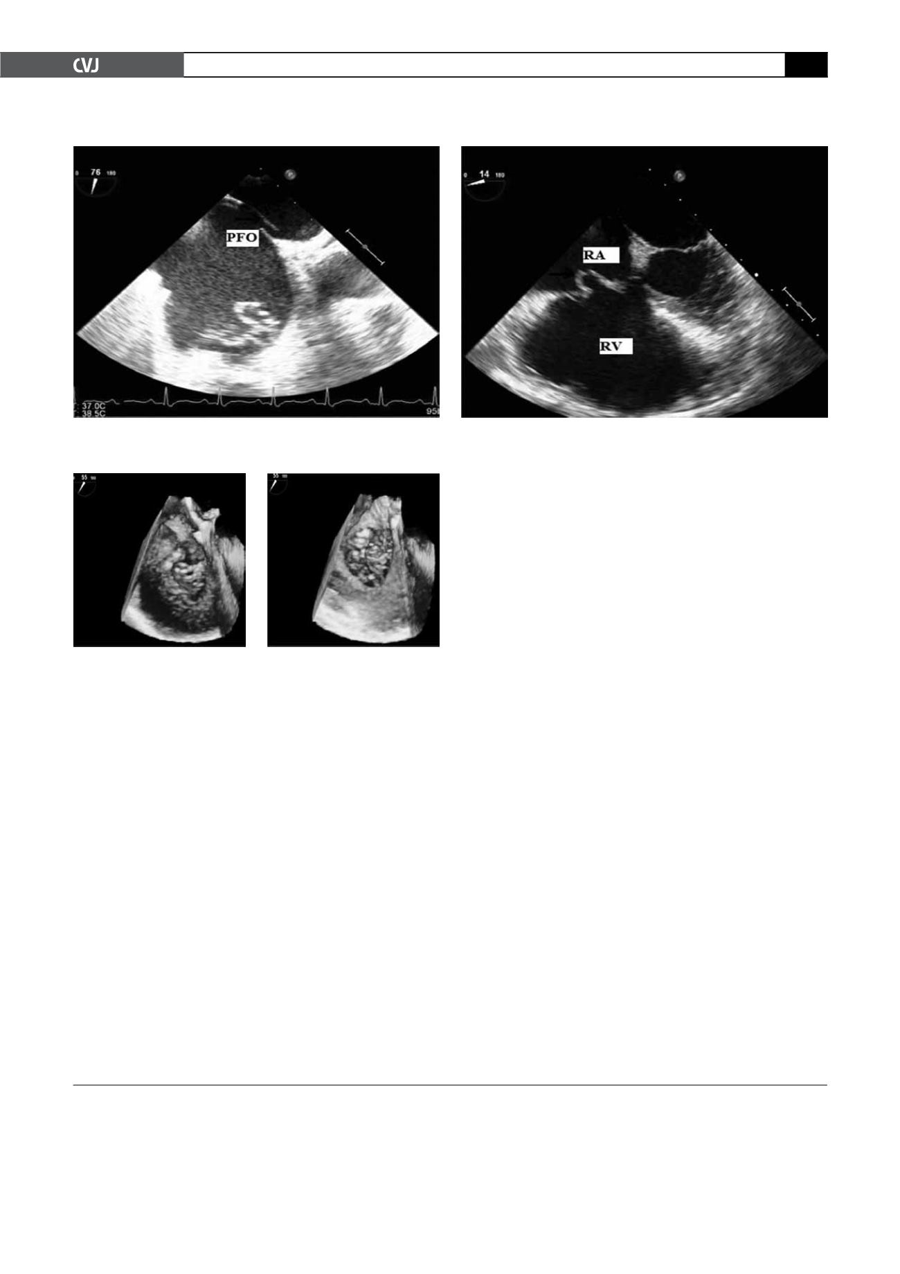
Fig. 2. A transoesophageal echocardiography (TEE) showing patent foramen ovale and a prolapse of the anterior
leaflet. RV: right ventricle, RA: right atrium, PFO: patent foramen ovale.
Fig. 3. Three-dimensional TEE.


