CARDIOVASCULAR JOURNAL OF AFRICA • Vol 23, No 10, November 2012
AFRICA
e5
Case Report
MRI finding of a papillary muscle cyst: a differential
diagnosis
THANDAZA SHAYINGCA, SAVVAS ANDRONIKOU, RENE TRUTER, EMILE REID
Abstract
Cystic lesions of the papillary muscle in the form of myxoma,
hydatid cyst, papillary fibroelastoma, blood-filled cysts and
endodermal heterotopia are rare causes of embolic stroke. In
view of the potential complications caused by these lesions,
surgery is often advocated but there is no consensus on
which patients qualify. We examined a differential diagnosis
of a papillary muscle cystic lesion in a patient presenting
with features of embolic disease and identified the imaging
features on MRI that directed management.
Keywords:
papillary muscle, myxoma, hydatid, fibroelastoma
Submitted 7/12/11, accepted 3/9/12
Cardiovasc J Afr
2012;
23
:
e5–e6
DOI: 10.5830/CVJA-2012-062
The papillary muscles are a specialised form of the trabeculae
carnae. They connect to the chordae tendinae, which attach to
the tricuspid valve in the right ventricle and the mitral valve
in the left ventricle, and act to prevent regurgitation by bracing
the atrio-ventricular valves against prolapse. Cystic lesions
of the papillary muscle in the form of myxoma, hydatid cyst,
papillary fibroelastoma, blood-filled cysts and endodermal
heterotopia are rare causes of embolic stroke.
1
In view of the
potential complications caused by these lesions, surgery is often
advocated but there is no consensus on which patients qualify.
We examined a differential diagnosis of a papillary muscle
cystic lesion in a patient presenting with features of embolic
disease, and identified the imaging features on MRI that directed
management.
Case report
A 50-year-old female investigated for recurrent transient
ischaemic attacks (TIA) underwent cardiac MRI following an
echocardiogram, which suggested a possible aneurysm of the
free wall of the left atrium. MRI (T1, STIR, T2 ciné gradient
protocols and post-contrast viability studies at different levels)
showed normal anatomical and ventriculo-atrial connections,
and normal concordance. MRI further demonstrated an abnormal
hyper-intense (STIR and T2 gradient echo) lesion in relation
to the posterior ventricular papillary muscle. The lesion was
cystic with subtle post-contrast enhancement. No involvement
of the surrounding ventricular wall or motion abnormality was
demonstrated.
A differential diagnosis of myxoma, fibroelastoma, and
even a papillary muscle pseudoaneurysm was considered.
The patient was subsequently referred for a transoesophageal
echocardiographic (TEE) assessment, which confirmed a 15
× 12-mm central lucent mass at the posterior-medial papillary
muscle, and mild central mitral valve regurgitation. The subtle
gadolinium enhancement made a neoplasm less likely, prompting
the adoption of a conservative approach to therapy, using
anticoagulation and a planned follow-up MRI after six months.
Discussion
The significance of the papillary muscle in cardiac function
was appreciated more than a century ago. Diseases of the
papillary muscle are mostly subtle, therefore causing a delay in
presentation, and they are mostly discovered at post mortem. In
this case report we present a differential diagnosis of a cystic
lesion in a patient who presented with TIA.
Myxomas are the most common primary tumours of the heart
and arise from the endocardium.
2
They tend to present as cavitary
gelatinous masses that arise adjacent to the fossa ovalis in the
left atrium and are typically pedunculated but can also arise in a
Department of Radiology, Faculty of Health Sciences,
University of the Witwatersrand, Johannesburg, South
Africa
THANDAZA SHAYINGCA, MD,
SAVVAS ANDRONIKOU, MD
Schnetler Corbett and Partners, Private radiology practice,
Cape Town, South Africa
RENE TRUTER, MD
Private practice, Cape Town, South Africa
EMILE REID, MD
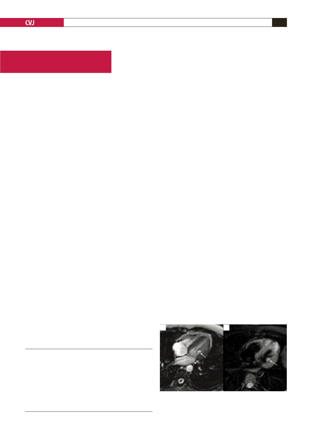
Fig. 1. Axial MRI demonstrating an abnormal hyperin-
tense (a) T2 gradient echo, and enhancing (b) corre-
sponding post-gadolinium lesion in relation to the poste-
rior ventricular papillary muscle (arrows).
a
b


