CARDIOVASCULAR JOURNAL OF AFRICA • Vol 24, No 8, September 2013
AFRICA
315
Patients were followed up in the third hour, seventh day, and
first and sixth month post-procedure. The patients registered a
pain score on a visual analogue scale (VAS) from 0 (no pain)
to 10 (worst pain ever). Also the ecchymosis scores of the
patients were scaled from 0 (light bruise) to 5 (critical bruise).
Clinical examination was performed at each visit and Doppler
ultrasonography was performed on the first and sixth months
postoperatively. The initial clinical result were assessed, GSV
diameters were measured and occlusion of GSV was confirmed
with ultrasonography.
Statistical analysis
Data analysis was performed using the SPSS for Windows 11.5
package program. The Shapiro Wilk test was used in order to
investigate whether the distribution of continous variables was
close to normal. Measurable parameters were expressed as mean
±
standard error of the mean. Intergroup comparisons were made
with the Student’s
t-
test or Mann-Whitney
U
-test, as appropiate,
for normally and non-normally distributed data, respectively.
Nominal variables were evaluated with Pearson’s chi-square test.
A
p
value
<
0.05 was considered statistically significant.
Results
A total of 344 patients were treated with endovenous
radiofrequency ablation. In group 1, tumescent anaesthesia
was given before the ablation procedure while in group 2, a
local hypothermia technique was used. The demographic data
of the patients are shown in Table 1. There was no significant
intergroup differences in terms of age, gender, body weight and
height, and body mass index (
p
>
0.05)
No statistically significant difference was found between the
groups in the pre-operative grey-scale measurements of GSV,
including diameter of the GSV in the SFJ, the diameter of the
GSV above the knee, the distance of the GSV from the skin
above the knee, and the length of the GSV that was to be ablated
(
p
>
0.05). However, the distance of the GSV from the skin in the
middle of the thigh was shorter in group 1 patients (
p
=
0.017)
(Table 2).
Mean ablation time was significantly lower in group 2
compared to group 1 (7.2 vs 18.9 min;
p
<
0.05). Skin burn and
paresthesia did not occur. Immediate occlusion rate was 100%
for both groups. All patients returned to normal activity within
two days.
All patients have reached the six-month follow-up point. We
recognised recanalisation in three patients in group 1 and two in
group 2 by Doppler ultrasound scanning. The primary closure
rate of group 1 was 98.2% and group 2 was 98.8% at six months
and there was no significant difference between the groups (
p
>
0.05). Endovenous heat-induced thrombosis and deep-vein
thrombosis were not observed in any of the patients.
The patients’ VAS scores were measured in the third hour,
seventh day, first month and sixth month (Table 3). We observed
there was no statistically significant difference between the pain
score for the patients who recieved tumescent anesthesia and
the patients on whom a local hypothermia and compression
technique was used (
p
>
0.05).
The patients’ ecchymoses scores were measured in the third
hour, seventh day, first month and sixth month (Table 4). There
was no significant difference between the groups in terms of
ecchymoses scores (
p
>
0.05).
At the one-month and six-month Doppler ultrasonography
follow up, the diameter of the GSV at the SFJ and above the
knee were measured. In both groups, GSV had decreased in
diameter but no significant difference was found between the
groups (Table 5).
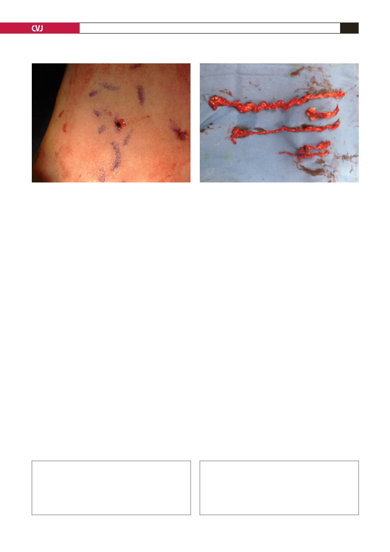
Fig. 2. Phlebectomy incision.
Fig. 3. Varicose veins.
TABLE 1. DEMOGRAPHIC DATA OF THE PATIENTS
Variables
Group 1 (
n
=
172)
Group 2 (
n
=
172)
p
-value
Age (years)
46.1
±
10.2
44.0
±
10.6
0.178
Gender (f/m)
114/58
130/42
0.179
Height (cm)
167.2
±
7.6
165.4
±
8.1
0.146
Weight (kg)
79.0
±
10.9
76.5
±
13.4
0.173
Body mass index (kg/m
2
)
28.3
±
3.8
28.0
±
4.7
0.592
TABLE 2. PRE-OPERATIVE GREY-SCALE MEASUREMENTS OF THE GSV
Measurement (cm)
Group 1 (
n
=
172) Group 2 (
n
=
172)
p
-value
Diameter of GSV above the knee 5.0 (4.0–12.1)
4.8 (4.0–11.9)
0.532
Diameter of GSV in SFJ
9.2 (6.2–23.0)
9.2 (4.9–23.0)
0.952
Length of GSV treated
44.0 (30.0–70.0) 42.5 (20.0–64.0) 0.147
GSV-skin distance above the knee 8.0 (1.3–28.8)
9.2 (1.0–32.2)
0.099
GSV-skin distance mid-thigh
11.3 (2.6–32.5)
14.0 (1.0–34.7)
0.017


