CARDIOVASCULAR JOURNAL OF AFRICA • Vol 23, No 1, February 2012
e8
AFRICA
CARDIOVASCULAR JOURNAL OF AFRICA • Vol 22, No 4, July/August
1
144
pheral and entrapment neuropathy, which was likely a chronic
demyelinating neuropathy or burnt-out variant of GBS.
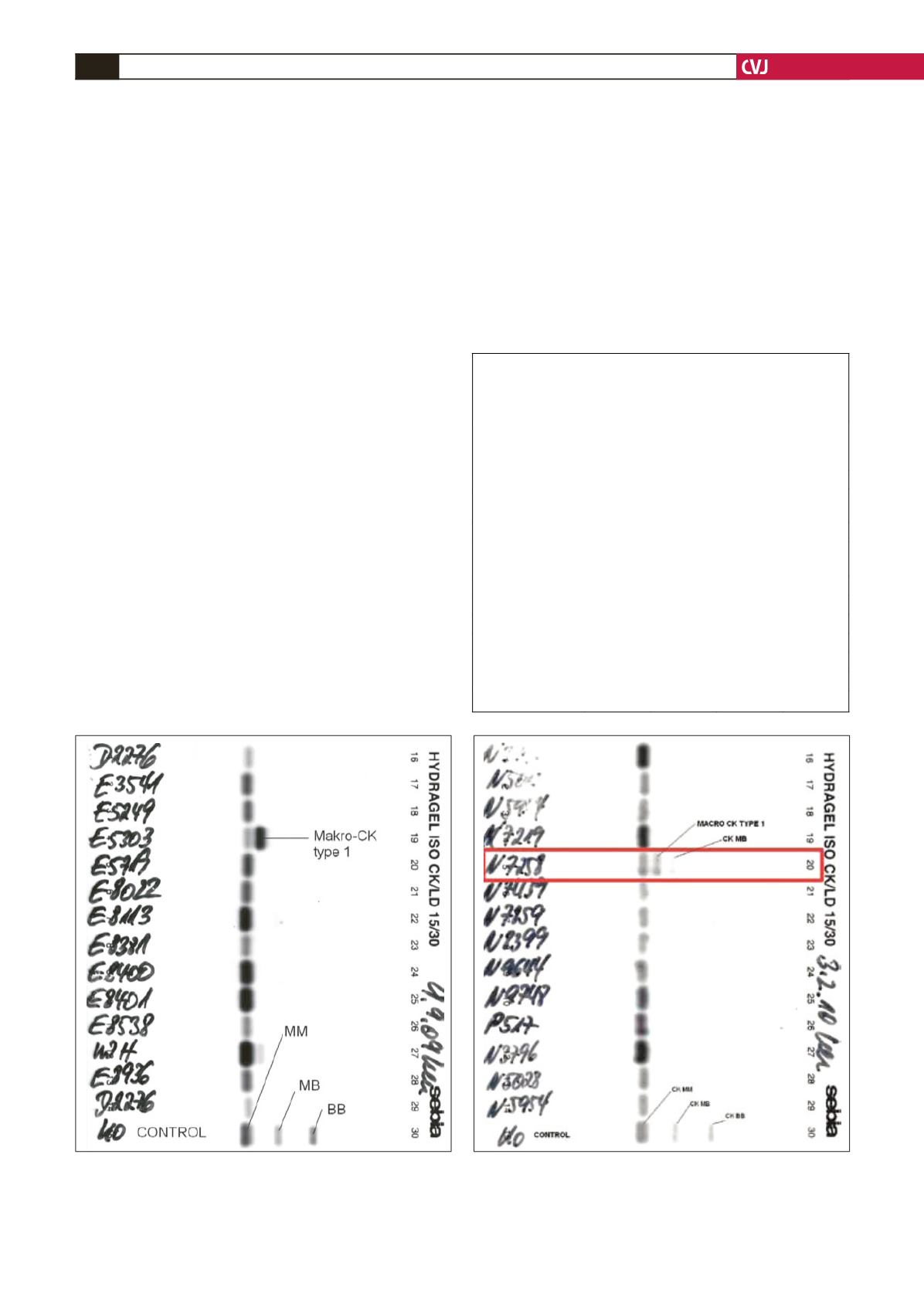
Electrophoresis of the serum creatine kinase enzyme
revealed the presence of macro CK type 1, as shown in Fig.
1. Electrophoresis was done using Hydragel
®
and Hydrasys
®
electrophoresis systems. Macro CK type 1 migrates between
CK-MM and CK-MB.
The patient was treated successfully and discharged home
after one month on physiotherapy and regular follow up.
Case report 2
A 60-year-old male patient was admitted in December 2009 and
discharged one week later with an acute myocardial infarction
for which successful thrombolysis was done. He had had symp-
toms of unstable angina in the two weeks prior to admission.
He was a known type 2 diabetic patient for 13 years, with
poorly controlled hypertension that had been diagnosed seven
years previously. He was also known to suffer from ulcerative
colitis, which was in remission on mesalamine. He was managed
for
Helicobacter pylori
-positive gastritis confirmed on biopsy
specimens. He had impaired vision in the right eye following
central retinal artery thrombosis in 2005.
On examination, the major findings were an apprehensive
and diaphoretic patient, with a blood pressure of 160/80 mmHg.
Jugular venous pressure was not elevated. He had right optic
nerve atrophy but no diabetic changes.
ECG showed an acute anterior ST-elevation myocardial infarc-
tion with an intra-ventricular conduction abnormality compara-
ble with incomplete right bundle branch block. Two-dimensional
echocardiogram revealed an ejection fraction of 50%, trivial
mitral regurgitation into a normal-sized left atrium and estimated
pulmonary artery pressure of 25 mmHg. Coronary angiography
at 48 hours post admission revealed bi-vessel disease (proximal
left anterior descending coronary artery and second obtuse
marginal disease).
The serum troponin I, total CK and CK-MB were as shown in
Table 2. As macro CK was suspected in this patient, isoenzyme
electrophoresis was done, as shown in Fig. 2.
Renal function tests done during and after admission revealed
a persistently elevated creatinine level (144 µmol/l) with an
estimated glomerular filtration rate of approximately 46 ml/min,
TABLE 2. TRENDS OF SERUM CK, CK-MBAND
TROPONIN I LEVELS FOR PATIENT 2.
THE NORMAL REFERENCEVALUES OF THE
ANALYTESARE INDICATED IN BRACKETS
CK (U/l)
(34–145)
CK-MB
(U/l)
(0–24)
Troponin I
(ng/ml)
(
<
0.4)
%CK-MB
(
<
6%)
Index day
316
252
0.18
79
Troponin I four
hours later
5.72
Day 2
279
245
6.23
88
Four hours later on
day 2
242
246
3.95
>
100
Day 3
179
229
1.92
>
100
Day 4
172
204
1.22
>
100
Day 5
169
228.6
0.75
>
100
Day 16
181
240
<
0.04
>
100
Day 34
235
254
<
0.04
>
100
Day 35
197
234.4
<
0.04
>
100
Day 36
203
252
<
0.04
Fig. 1. Electrophoresis of serum creatine kinase isoen-
zymes showing the presence of macro CK type 1 in our
patient; patient result is highlighted.
Fig. 2. Electrophoresis of serum creatine kinase isoen-
zymes showing the presence of macro CK type 1 in our
patient; patient result is highlighted.
CARDIOVASCULAR JOURNAL OF AFRICA • Vol 22, No 4, July/August 2011
144
AFRICA
pheral and entrapment neuropathy, which was lik ly a chronic
demyelinating neuropathy or burnt-out variant of GBS.
Electrophoresis of the serum creatine kinase enzyme
revealed the presence of macro CK type 1, as shown in Fig.
1. Electrophoresis was done using Hydragel
®
and Hydrasys
®
electrophoresis systems. Macro CK type 1 migrates between
CK-MM and CK-MB.
The patient was treated successfully and discharged home
after one month on physiotherapy and regular follow up.
Case report 2
A 60-year-old male patient was admitted in December 2009 and
discharged one week later with an acute myocardial infarction
for which successful thrombolysis was done. He had had symp-
toms of unstable angina in the two weeks prior to admission.
He was a known type 2 di betic patient for 13 years, with
poorly controlled hypertension tha had b en diagnos d seven
years previously. He was also known to suffer from ulcerative
colitis, which was in remission on me alamine. He was managed
for
Helicobacter pylori
-positive gastritis confirmed on biopsy
specimens. He had impaired vision in the right eye following
central retinal artery thrombosis in 2005.
On examination, the major findings were an apprehensive
and diaphoretic patient, with a blood pressure of 160/80 mmHg.
Jugular venous pressure was not elevated. He had right optic
nerve atrophy but no diabetic changes.
ECG showed an acute anterior ST-elevation myocardial infarc-
tion with an intra-ventricular conduction abnormality compara-
ble with incomplete right bundle branch block. Two-dimensional
echocardiogram revealed an ejection fraction of 50%, trivial
mitral regurgitation into a normal-sized left atrium and estimated
pulmonary artery pressure f 25 mmHg. Coronary angiography
at 48 hours post admission revealed bi-vessel disease (proximal
left anterior descending coronary artery and second obtuse
marginal disease).
The serum troponin I, total CK and CK-MB were as shown in
Table 2. As macro CK was suspected in this patient, isoenzyme
electrophoresis was done, as shown in Fig. 2.
Renal function tests done during and after admission revealed
a persistently elevated creatinine level (144 µmol/l) with an
estimated glomerular filtration rate of approximately 46 ml/min,
TABLE 2. TRENDS OF SERUM CK, CK-MBAND
TROPONIN I LEVELS FOR PATIENT 2.
THE NORMAL REFERENCEVALUES OF THE
ANALYTESARE INDI ATED IN BRACKETS
CK (U/l)
(34–145)
CK-MB
(U/l)
(0–24)
Troponin I
(ng/ml)
(
<
0.4)
%CK-MB
(
<
6%)
Index day
316
252
0.18
79
Troponin I four
hours later
5.72
Day 2
279
245
6.23
88
Four hours later on
day 2
242
246
3.95
>
100
Day 3
179
229
1.92
>
100
Day 4
172
204
1.22
>
100
Day 5
169
228.6
0.75
>
100
Day 16
181
240
<
0.04
>
100
Day 34
235
254
<
0.04
>
100
Day 35
197
234.4
<
0.04
>
100
Day 36
203
252
<
0.04
Fig. 1. Electrophoresis of serum creatine kinase isoen-
zymes showing the presence of macro CK type 1 in our
patient; patient result is highlighted.
Fig. 2. Electrophoresis of serum creatine kinase isoen-
zymes showing the presence of macro CK type 1 in our
patient; patient result is highlighted.
CARDIOVASCULAR JOURNAL OF AFRICA • Vol 22, No 4, July/August 2011
144
AFRICA
pheral and entrapment neuropathy, which was likely a chronic
d myelinating neuropathy or burnt-out variant of GBS.
Electrophore is of the serum creatin kinase enzyme
revealed the presence of macr
e , as shown in Fig.
1. Electrophoresis was done using Hydragel
®
and Hydrasys
®
electro horesis systems. Macro CK t pe 1 m grates between
CK-MM and CK-MB.
The patient was treated successfully and discharged home
after one month on physiotherapy and regular follow up.
Case report 2
A 60-year-old male patient was admitted in December 2009 and
discharged one week later with an acute myocardial infarction
for which successful thrombolysis was done. He had had symp-
toms of unstable angina in the two weeks prior to admission.
He was a known type 2 diabetic patient for 13 years, with
poorly controlled hypertension that had been diagnosed seven
years previously. He was also known to suffer from ulcerative
colitis, which was in remission on mesalamine. He was managed
for
Helicobacter pylori
-positive gastritis confirmed on biopsy
specimens. He had impaired vision in the right eye following
central retinal artery thro b sis i 2005.
On exam nation, he major fin ings were an apprehensive
and diaphoretic patient, with a blood pressure of 160/80 mmHg.
Jugula venous pressure was not elevated. He had right optic
nerve atr phy but no diabe ic chang s.
ECG showed an acute anterior ST-elevatio myocardial infarc-
tion with an intra-ventricular conduction abnormality compara-
ble with inco plete right bundle branch block. Two-dimensional
echocardiogram revealed an ejection fraction of 50%, trivial
mitral regurgitation into a normal-sized left atrium and estimated
pulmonary artery pressure of 25 mmHg. Coronary angiography
at 48 hours post admission revealed bi-vessel disease (proximal
left ant rio descending coronary artery and second obtuse
marginal disease).
The serum troponin I, total CK and CK-MB were as shown in
Table 2. As ma ro CK was suspected in this patient, oenzyme
electrophoresis was done, as shown in Fig. 2.
Renal function tests done duri g and after admission revealed
a persistently elevated creatinine level (144 µmol/l) with an
estimated glomerular filtration rate of approximately 46 ml/min,
TABL 2. T ENDS OF S UM CK, CK-MBAND
TROPONIN I LEVELS FOR PATIE T 2.
THE NORMAL REFERENCEVALUES OF THE
ANALYTESARE INDICATED IN BRACKETS
CK (U/l)
(34–145)
CK-MB
(U/l)
(0–24)
Troponin I
(ng/ml)
(
<
0.4)
%CK-MB
(
<
6%)
Index day
316
252
0.18
79
Troponin I four
hours later
5.72
Day 2
279
245
6.23
88
Four hours later on
day 2
242
246
3.95
>
100
Day 3
179
229
1.92
>
100
Day 4
172
204
1.22
>
100
Day 5
169
228.6
0.75
>
100
Day 16
181
240
<
0.04
>
100
Day 34
235
254
<
0.04
>
100
Day 35
197
234.4
<
0.04
>
100
Day 36
203
252
<
0.04
Fig. 1. Electrophoresis of serum creatine kinase isoen-
zymes showing th presence of macro CK type 1 in ur
patient; pat ent result i highlighted.
Fig. 2. Electrophoresis of serum creatine kinase isoen-
zymes showing the presence of macro CK type 1 in our
patient; patient result is highlighted.


