


 44 / 72

CARDIOVASCULAR JOURNAL OF AFRICA • Volume 27, No 2, March/April 2016
98
AFRICA
Echocardiography
Echocardiography is an imaging modality that uses high-
frequency (2–10 MHz) sound waves to image cardiac structures
and to give reproducible information about cardiac structure and
function. Ultrasound is produced when a piezo-electric crystal,
mounted in a transducer, is stimulated by an electric current.
30
Ultrasound waves are not audible and are harmless to tissue
at the intensities used in diagnostic imaging. The passage of
sound waves depends on the acoustic impedance of tissues.
Most ultrasound waves pass through tissues to deeper structures
further from the surface, but reflected sound returns to strike
the crystal, deforming it and producing electric signals, which
correspond to the degree of deformation.
30
This electrical
information is transformed so it can be displayed on a cathode-
ray tube as pulses of light. Due to the speed of sound within the
body being relatively constant, the depth of the tissue interface
can be calculated, and reflected echoes are displayed on the
screen on a depth scale.
31
Blood reflects little sound and appears relatively black/hypo-
echoic compared with the myocardium, which reflects more of
the ultrasound and appears relatively white/hyperechoic. The
heart valves are even more echogenic. Neither bone nor air is
a good transmission medium for ultrasound waves; therefore
as the heart is surrounded by lung and the bony cage of the
thoracic cavity, the ultrasound beam must be aimed through
specific gaps, known as acoustic windows (e.g. parasternal,
apical, subcostal and suprasternal), to produce images of the
heart and vasculature.
31
Given the lack of ionising radiation,
echocardiography is an attractive first-line investigation for most
forms of CVD encountered in pregnancy.
M-mode and two-dimensional (2D) echocardiography provide
real-time imaging of heart structures throughout the cardiac
cycle; more recently, three-dimensional (3D) echocardiography
has been developed.
32
Doppler echocardiography provides
information on blood movement inside cardiac structures and
on the haemodynamics
33
(Fig. 2). Tissue Doppler imaging (TDI)
provides information about movement of cardiac structures.
33
The relationship between the dynamics of cardiac structures and
the haemodynamics of the blood inside these structures provides
information about cardiac diastolic and systolic function.
33
Echocardiography is continuously evolving and constantly being
augmented by newer modalities, such as tissue harmonics, speckle
tracking, tissue Doppler strain and tissue characterisation.
34
To date, there have been no reports of documented adverse
foetal effects from diagnostic ultrasound procedures, including
duplex Doppler imaging.
1
There are no contra-indications to
echocardiography during pregnancy, and ultrasound is preferred
over X-ray as the primary method of foetal imaging during
pregnancy.
35
Energy exposure from ultrasonography has been
arbitrarily limited to 94 mW/cm
2
by the US Food and Drug
Administration (FDA).
36
Doppler and colour echocardiographic scans work by
concentrating a beam of sound in a small area, and therefore
can cause heating of local tissues if held in the same place for
a long time.
37
Most scans automatically reduce the power of the
ultrasound beam when Doppler is used, to decrease the intensity.
Nowadays most echocardiographic machines have a low thermal
index and so pose very little risk. Dobutamine is favoured in
pregnancy over adenosine for stress echocardiography.
38
Cardiovascular computed tomography (CCT)
Computed tomography (CT) is a diagnostic imaging procedure
that uses X-rays to demonstrate cross-sectional images of
the body acquired in different orthogonal planes. The cross-
sections or slices are reconstructed from the measurements of
attenuation coefficients of X-ray beams in the volume of the
object studied.
39
The fundamental principle of CT is premised
on tissue density traversed by the X-ray beam, which can be
calculated from the attenuation coefficient. In other words,
CT permits reconstruction of tissue density by 2D sections
perpendicular to the axis of the acquisition system. Unlike X-ray
radiography, the detectors of the CT scanner do not produce
an image, but rather measure the transmission of a thin beam
(1–10 mm) of X-rays through a full scan of the body, and the
image of that section is taken from different angles, allowing
retrieval of information on the depth of the tissues imaged.
40
Complex mathematical algorithms are used to construct an
image from the raw data; a typical CT image is composed of
512 rows, each of 512 pixels, i.e. a square matrix of 512 × 512 =
262 144 pixels (one for each voxel). A typical CCT study gives
0.06 to 0.09 rad.
41
Similarly, the effective radiation dose for CT
pulmonary angiogram (CTPA) protocols is generally between
2.2 and 7 mSv (0.02–0.07 rad).
42
Often, for CCT and CTPA, the imaging field of view includes
the lungs and breasts; the radiation dose can be reduced by
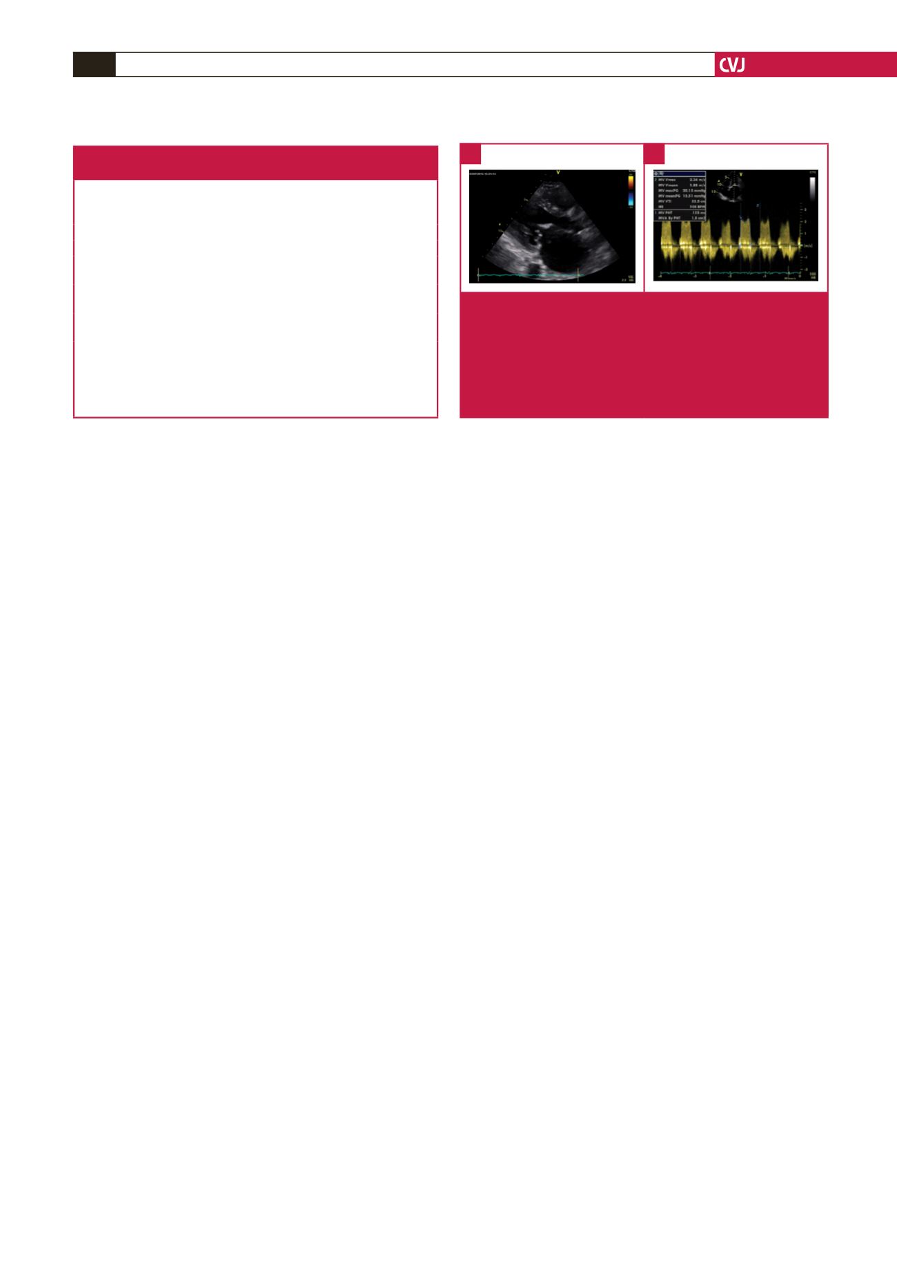
Fig. 2.
Echocardiography in a pregnant woman with mitral
stenosis. (A) parasternal long-axis view showing a
deformed, calcified and restricted mitral valve with a
classic ‘hockey-stick’ deformity of the anterior mitral
valve leaflet and a dilated left atrium. (B) continuous-
wave Doppler trace showing a mean gradient of 15.5
mmHg, indicating severe mitral stenosis.
A
B
Table 3. Approaches to minimising foetal radiation during
cardiovascular imaging in pregnancy
Restricting the X-ray beam size to as small as is necessary
Choosing the direction of the primary beam so that it is as far away from
the foetus as possible
Ensuring that the overall exposure time is as short as possible
Selecting appropriate exposure factors
Defer abdominal examinations if possible; imaging examinations of the
thorax are associated with negligible risks to the conceptus
Whenever possible, ultrasound is the preferred modality for abdominal
imaging in pregnancy
Magnetic resonance imaging is emerging as an alternative in centres where
it is widely available
Using a lead apron on the table to shield any primary beam from the X-ray
tube reaching the foetus
Calculations of dose by a knowledgeable medical physicist if there is concern
The radiation dose should be kept as low as reasonably achievable
(ALARA principle)