CARDIOVASCULAR JOURNAL OF AFRICA • Vol 23, No 5, June 2012
AFRICA
e3
Case Report
The dangerous fifth chamber: congenital left atrial
appendage aneurysm
KURSAT M Tigen, CEM Dogan, AHMET Guler, SUZAN Hatipoglu, MEHMET Yanartas, CEVAT Kirma
Abstract
Aneurysms of the left atrial appendage are extremely rare.
Enlargement of the left atrial appendage can be congeni-
tal or acquired. Dysplasia of the left atrial muscles leads
to congenital left atrial appendage aneurysm and usually
presents as atrial tachyarrhythmia or embolic events in
the second or third decade of life. We report a case of an
asymptomatic 12-year-old child with a congenital left atrial
appendage aneurysm. Transthoracic and transoesophageal
echocardiography demonstrated a large left atrial appendage
aneurysm without thrombus or spontaneous echo-contrast.
The patient was successfully treated with surgical resection
of the aneurysm.
Keywords:
congenital, left atrial appendage aneurysm
Submitted 7/2/11, accepted 6/9/11
Cardiovasc J Afr
2012;
23
: e3–e4
DOI: 10.5830/CVJA-2011-051
Case report
A 12-year-old female without symptoms and previous history
of disease was referred to our clinic because of a chest X-ray
demonstrating a prominent convexity of the left upper heart
border in the position of the left atrial appendage (LAA). The
physical examination was normal and an electrocardiograph
(ECG) showed normal sinus rhythm. Laboratory findings were
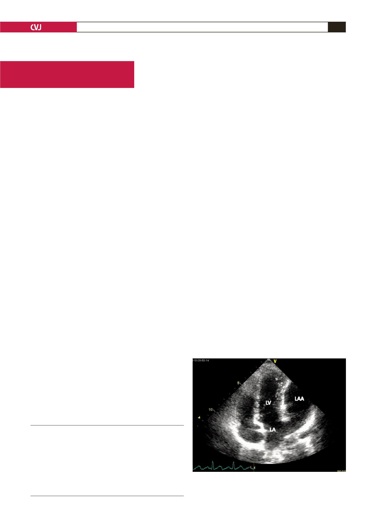
within the normal range. Transthoracic echocardiography (TTE)
revealed a huge LAA aneurysm resembling a fifth cardiac
chamber and normal ventricular and valvular function (Fig. 1).
For better determination of the aneurysm, transoesophageal
echocardiography (TEE) was performed. This revealed a 6.2 ×
4.4-cm LAA aneurysm with a left atrial connection. There was
no spontaneous echo-contrast or thrombus in the LAA and left
atrium, and there was dynamic blood flow within the aneurysm
(Fig. 2).
Because of the potential risk of embolism, the patient was
referred to surgery for resection of the aneurysm. Following a
median sternotomy, surgical exploration confirmed the diagnosis
of an LAA aneurysm (Fig. 3). The aneurysm was resected
without extracorporeal circulation. The anatomical features of
the resected LAA were consistent with the echocardiographic
images. The patient’s postoperative course was uneventful.
Discussion
Aneurysm of the LAA is a very rare condition and can be
categorised into two groups; congenital or acquired. Acquired
LAA aneurysms are associated with increased left atrial pressure
as a result of rheumatic valve disease or mitral regurgitation. The
cause of congenital aneurysms of the LAA is dysplasia of the
pectineal muscles.
1
Symptoms associated with aneurysms of the LAA are
supraventricular tachyarrhythmia, such as atrial fibrillation or
flutter and paroxysmal atrial tachycardia, embolic events, heart
failure and angina pectoris. Atrial tachyarrhythmia, which is
the most common cause for presentation, could be explained
by irritation of the enlarged LAA or congenital defects of the
conduction system. Embolic events such as cerebrovascular
infarcts are related to stasis caused by the increase in LAA
volume and subsequent thrombus formation. More rarely in
Department of Cardiology, Kartal Kosuyolu Heart Education
and Research Hospital, Istanbul, Turkey
KURSAT M TIGEN, MD
CEM DOGAN, MD
AHMET GULER, MD,
Suzan Hatipoglu, MD
CEVAT KIRMA, MD
Department of Cardiovascular Surgery, Kartal Kosuyolu
Heart Education and Research Hospital, Istanbul, Turkey
MEHMET YANARTAS, MD
Fig. 1. Apical four-chamber view of TTE demonstrating a
large LAA aneurysm resembling a fifth cardiac chamber.
LA: left atrium, LV: left ventricle, LAA: left atrial append-
age.


