CARDIOVASCULAR JOURNAL OF AFRICA • Vol 23, No 5, June 2012
AFRICA
e5
Case Report
Corrected transposition of the great arteries with
previously unreported cardiac anomalies
AHMET KAYA, IBRAHIM HALIL TANBOGA, MUSTAFA KURT, TURGAY IŞIK, MESUT OZGOKCE,
SELIM TOPÇU, ENBIYA AKSAKAL
Abstract
The corrected transposition of the great arteries is a rare
congenital cardiac anomaly characterised by atrio-ventricu-
lar and ventriculo-arterial discordance and is related to the
largest incidence of cardiological complications. We report
on a 40-year-old woman with congenitally corrected trans-
position of the great arteries, situs inversus, atrial septal
defect, pulmonary stenosis, right arcus aorta and coronary
artery anomalies.
Keywords:
corrected transposition, congenital heart disease,
cardiovascular imaging
Submitted 16/4/11, accepted 6/9/11
Cardiovasc J Afr
2012;
23
: e5–e7
DOI: 10.5830/CVJA-2011-049
Congenital cardiac anomalies are present in between
approximately 3.7 and eight of every 1 000 live-birth infants.
The corrected transposition of the great arteries (CTGA),
characterised by atrioventricular and ventriculo-arterial
discordance and normal atrial situs, occurs in approximately 0.5
to 1.4% of all congenital cardiac diseases.
1
Most patients have one or more associated cardiac anomalies,
and the presence or absence of these anomalies significantly alters
the natural history.
2
Peri-membranous ventricular septal defect
(VSD) and pulmonary stenosis (PS), which may result from an
aneurysm of the interventricular septum, an associated fibrous
tissue tag, or a discrete ring of tissue in the subvalvular area,
are the most common associated defects.
3
VSD occurs in 70%
of patients and PS in 40%.
2
Systemic ventricular dysfunction,
tricuspid regurgitation, heart blocks and arrhythmias are life-
threatening late complications in this patient group.
4
In the medical literature, there is no evidence of any patient
with corrected transposition combined with atrial septal defect
(ASD), pulmonary stenosis, situs inversus totalis, right arcus
aorta and coronary artery anomalies. We report here, for the first
time, a case of CTGA with all of these anomalies.
Case report
A 40-year-old woman was admitted to the cardiology clinic
with chest pain and worsening dyspnoea. She had no history
of cardiac disorder or risk factors for coronary artery disease.
Her blood pressure and pulse rate were normal at 125/80
mmHg and 60 beats/min, respectively. Auscultation revealed a
grade III holosystolic murmur, which was best heard at the left
sternal border. There was no diastolic murmur. The laboratory
examinations (blood count, biochemistry) were within normal
ranges, and electrocardiography (ECG) demonstrated regular
sinus rhythm.
Transthoracic echocardiography (TTE) showed atrio-
ventricular and ventriculo-arterial discordance and also revealed
that the aorta was on the right side, anteriorly located. The final
diagnosis was congenitally corrected transposition of the great
arteries. TTE further revealed a normal systemic (morphological
right) ventricular ejection fraction of 60%, and a normal
Department of Cardiology, Erzurum Regional Education and
Research Hospital Erzurum, Turkey
AHMET KAYA, MD,
IBRAHIM HALIL TANBOGA, MD
MUSTAFA KURT, MD
TURGAY IŞIK, MD
Department of Radiology, Medical Faculty, Atatürk
University Erzurum, Turkey
MESUT OZGOKCE, MD
Department of Cardiology, Medical Faculty, Atatürk
University Erzurum, Turkey
SELIM TOPÇU, MD
ENBIYA AKSAKAL, MD
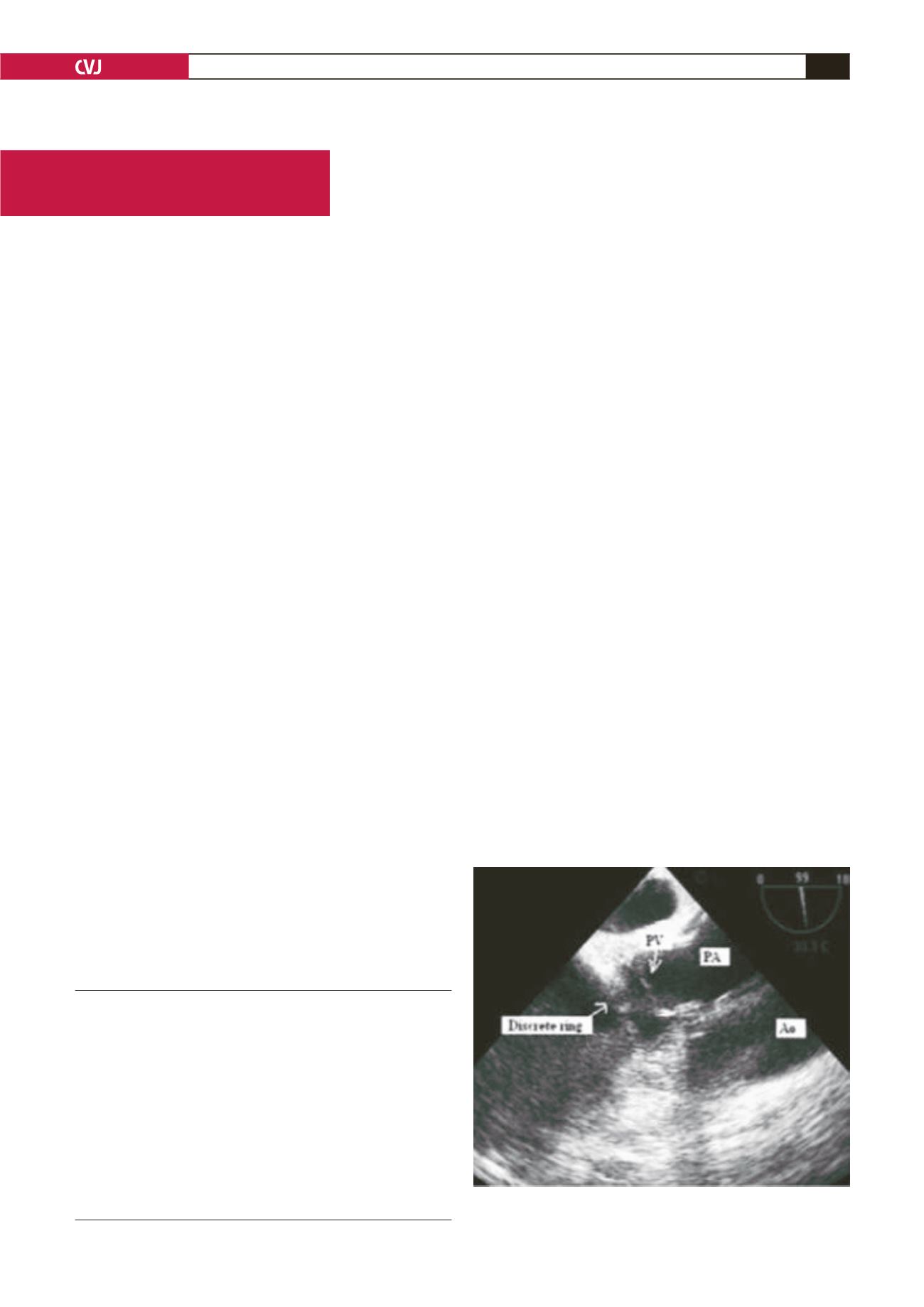
Fig. 1. TEE showing a discrete ring. PV: pulmonary valve,
Ao: aorta, PA: pulmonary artery.


