CARDIOVASCULAR JOURNAL OF AFRICA • Vol 23, No 5, June 2012
e6
AFRICA
pulmonary (morphological left) ventricular ejection fraction
of 55%. There was ventricular inversion with anatomical
preservation of the atrial and venous drainages, and the right
ventricle (RV) was dilated.
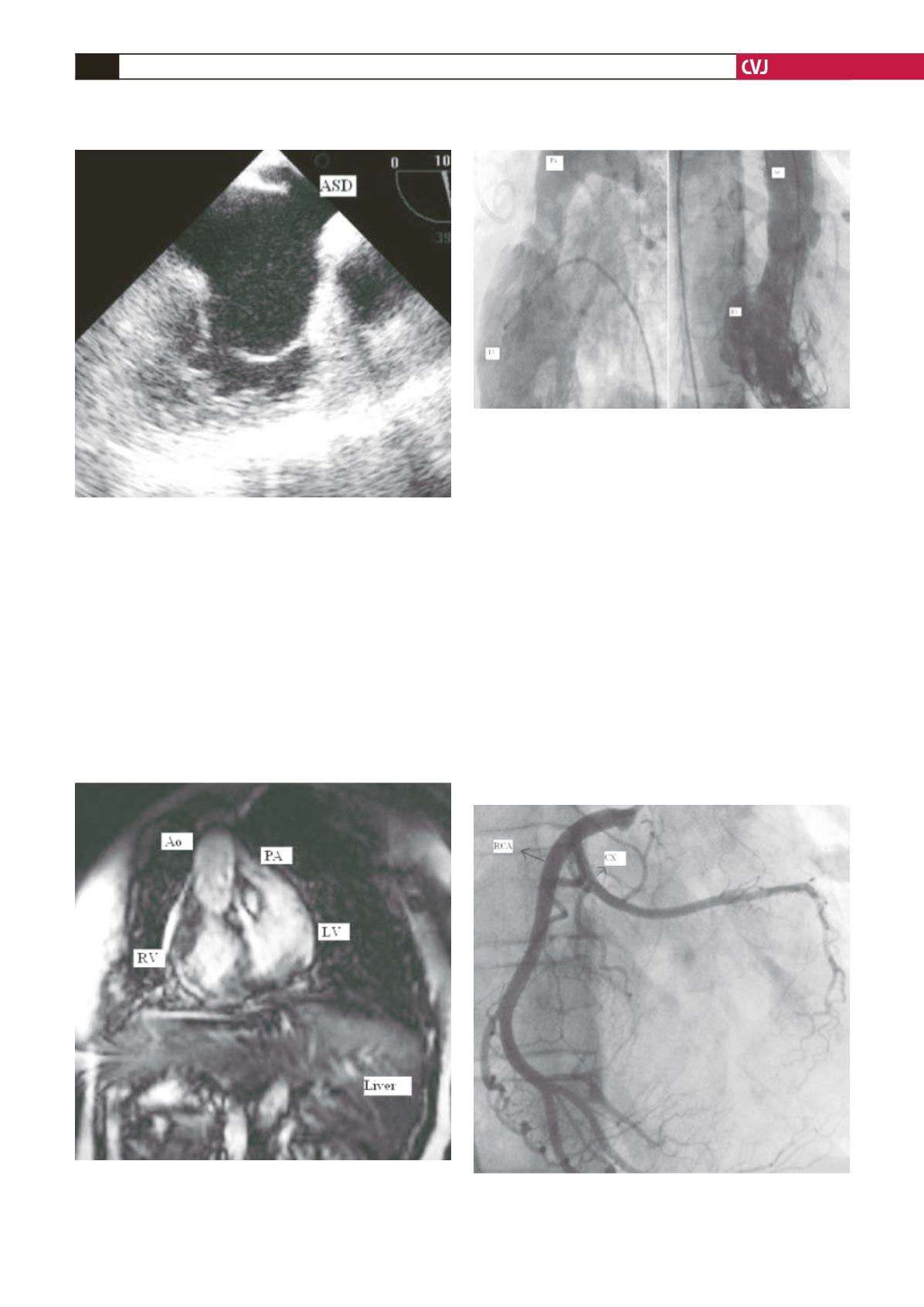
Transoesophageal echocardiography (TEE) showed there
was significant pulmonary obstruction associated with a
discrete ring of tissue in the subvalvular area (Fig. 1) and that
the transpulmonary gradient pressure was high (81 mmHg).
Additionally, there was an inter-atrial septal defect associated
with intra-cardiac left-to-right shunting (Fig. 2).
Cardiac magnetic resonance imaging (MRI) was performed
to further evaluate the cardiac morphology and coronary
anatomy. The results of MRI corroborated the findings on
echocardiography (Fig. 3).
The patient underwent right and left heart catheterisation,
which showed increased right-side pressures (130 mmHg).
Systemic ventricular end-diastolic pressures were normal.
Systemic ventriculography revealed a heavily trabeculated
ventricular chamber with an outflow tract leading to the aortic
valve, which is an indication of a morphological right ventricle
(Fig. 4) and D-transposition.
There was no angiographic evidence of significant systemic
atrio-ventricular valvular regurgitation, but there was intra-
cardiac shunting on the inter-atrial septum. Selective coronary
angiography revealed the circumflex artery originating from the
right coronary artery, and a normal course of the left anterior
descending and right coronary arteries (Fig. 5).
Fig. 5. Circumflex artery originating from the right coro-
nary artery. CX: circumflex artery, RCA: right coronary
artery.
Fig. 4. Left and right heart catheterisation. Right ventricu-
lography showing prominent trabeculations, which are
characteristic of a morphological right ventricle. RV: right
ventricle, Ao: aorta.
Fig. 2. TEE showing inter-atrial septal defect with intra-
cardiac shunting. ASD: atrial septal defect.
Fig. 3. Cardiac magnetic resonance imaging showing
CTGA, right arcus aorta and situs inversus. RV: right
ventricle, LV: left ventricle, Ao: aorta, PA: pulmonary
artery.


