CARDIOVASCULAR JOURNAL OF AFRICA • Vol 23, No 5, June 2012
e8
AFRICA
Case Report
Rocking mitral annuloplasty ring
PRASHANTH PANDURANGA, MOHAMMED K MUKHAINI
Abstract
Dehiscence of a mitral annuloplasty ring is a rare occur-
rence. We present a young patient with long-standing gross
dehiscence of a Duran annuloplasty ring secondary to suture
dehiscence, occurring three years after mitral valve surgery.
It was detected by transthoracic echocardiography. This case
emphasises the importance of clinical and echocardiographic
follow-up examinations after mitral valve surgery to detect
any unexpected complications.
Keywords:
Duran annuloplasty ring, dehiscence, mitral regur-
gitation
Submitted 16/7/10, accepted 12/9/11
Cardiovasc J Afr
2012;
23
: e8–e10
DOI: 10.5830/CVJA-2011-055
Dehiscence of a mitral annuloplasty ring is rare. We describe
a young patient with long-standing dehiscence of a Duran
annuloplasty ring, which on transthoracic echocardiography was
seen rocking.
Case report
A 21-year-old man with mitral valve prolapse and severe
symptomatic mitral regurgitation (MR) had undergone minimally
invasive mitral valve repair three years earlier. Through an upper-J
mini-sternotomy into the fourth right intercostal space, under
standard moderate hypothermic cardiopulmonary bypass, he
had undergone anterior leaflet (A2 segment) chordal shortening,
followed by the placement of a 31-mm Duran flexible mitral
ring, using interrupted sutures.
Post-operatively as well as at his first annual echocardiographic
examination, he showed no MR. He was followed up in the
cardiology clinic and was doing well, with no symptoms. On
cardiac auscultation, however, there was the appearance of a
pansystolic murmur, which had not been noted after the surgery.
There was no history of endocarditis or trauma.
Transthoracic and transoesophageal echocardiographic
studies revealed a rocking Duran annuloplasty ring in the left
atrium, with severe eccentric, posterolateral-directed MR (Figs
1, 2). The Duran ring had completely detached from the mitral
annulus except at the lateral commissural area and was flail
inside the left atrium. There was severe A2 prolapse with no
chordal rupture seen. His left ventricle measured 62 mm in
diastole and 44 mm in systole, with an ejection fraction of 60%.
The patient was repeatedly advised to have the mitral valve
surgery redone but he declined. He was doing well without any
symptoms or haemolysis at the two-year follow up.
Discussion
Mitral valve annuloplasty with flexible rings is a safe and stable
reconstructive procedure in which preservation of spatial motility
and configuration of the annulus allows a more physiologically
natural valve repair with improvement of ventricular function.
In one study, five-year significant MR-free survival was 75.1
±
4.6% for the group of patients with Carpentier rings and 82.4
±
4.5% for the group with Duran rings, with no significant
difference.
1
In another study using Duran rings, freedom from
re-operation at seven years was 98% for both ischaemic and
degenerative MR.
2
Post mitral valve repair, early recurrent MR is usually
procedure related [incomplete initial repair, suture dehiscence
Department of Cardiology, Royal Hospital, Muscat,
Sultanate of Oman
PRASHANTH PANDURANGA, MD, MRCP (UK), prashanthp_69@
yahoo.co.in
MOHAMMED K MUKHAINI, MD, FRCPC, FACC
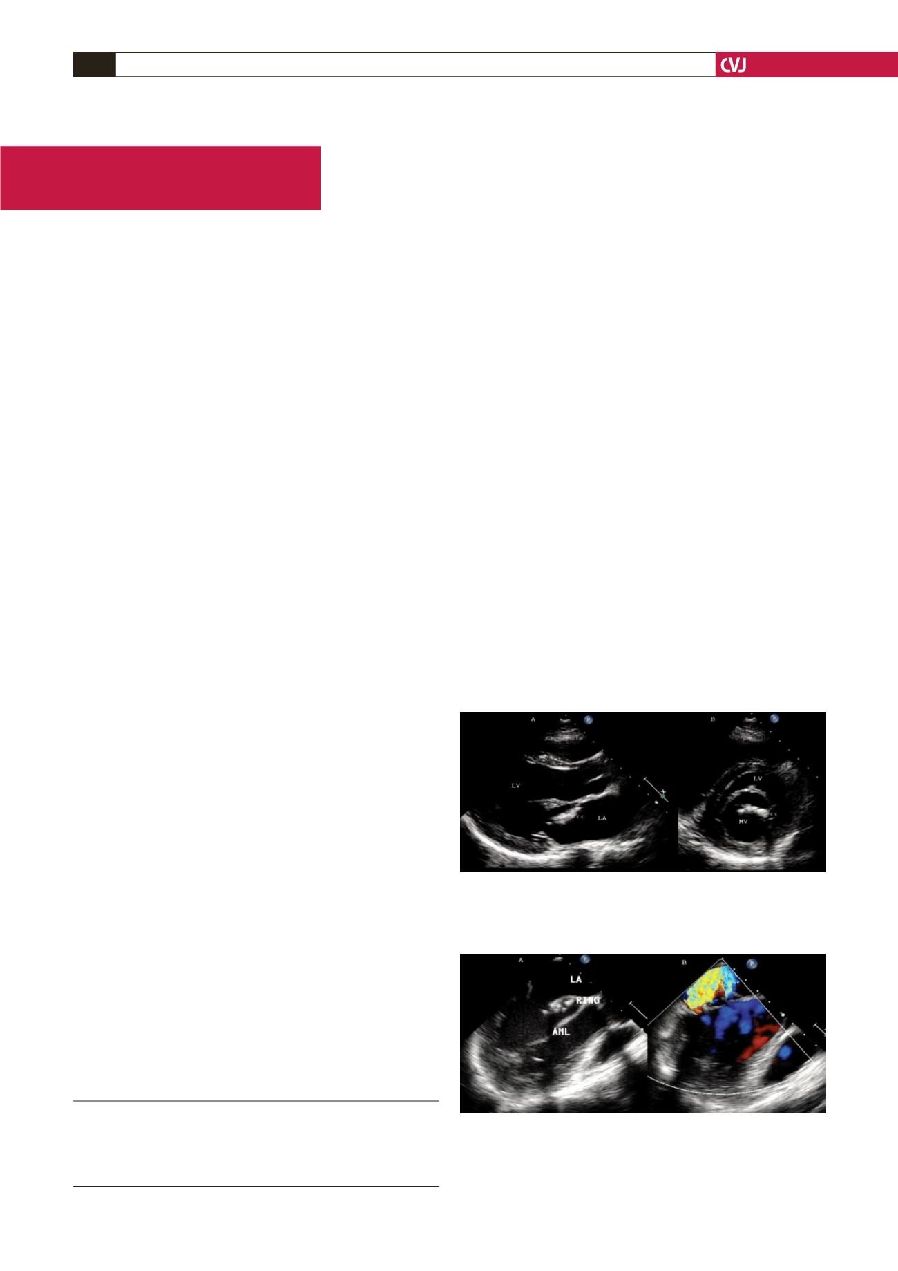
Fig. 1. Transthoracic echocardiography in the parasternal
long-axis (A) and short-axis (B) views showing gross
dehiscence of the Duran mitral annuloplasty ring (arrow-
heads). LA, left atrium; LV, left ventricle; MV, mitral valve.
Fig. 2. Transoesophageal echocardiography showing
dehiscence of the Duran mitral annuloplasty ring along
with the disrupted sutures (A). Colour Doppler (B) show-
ing the severe eccentric mitral regurgitation jet. LA, left
atrium; AML, anterior mitral leaflet.


