CARDIOVASCULAR JOURNAL OF AFRICA • Vol 23, No 5, June 2012
e4
AFRICA
the literature, there are also reports of patients presenting with
positional hypotension and tachycardia.
2
An echocardiographic image of an LAA aneurysm could be
confused with cystic tumours of the mediastinum, pericardial
cysts, herniation of the left atrium through a pericardial defect,
anomalous pulmonary venous drainage, and secondary causes
of LAA enlargement.
3-5
TEE, computed tomography and cardiac
magnetic resonance imaging provide detailed information on the
structure and composition of the LAA aneurysm.
LAA aneurysms should be treated even if the patient is
asymptomatic because they could be the source of arrhythmias
and life-threatening thromboembolic complications. The surgical
treatment of congenital aneurysm of the LAA is resection with or
without extracorporeal circulation.
Conclusion
A left atrial appendage aneurysm is a rare but potentially
dangerous entity. Because of the risk of embolic complications,
resection is advised when possible.
References
1.
Chowdhury UK, Seth S, Govindappa R, Jagia P, Malhotra P. Congenital
left atrial appendage aneurysm: a case report and brief review of litera-
ture.
Heart Lung Circ
2009;
18
(6): 412‒416.
2.
Zhang PF, Zhang M, Zhang W, Yao GH, Wu SM, Zhang Y. Giant aneu-
rysm of the left atrial appendage: detected by real-time 3-dimensional
echocardiography.
Tex Heart Inst J
2010;
37
(1): 129‒130.
3.
Foale RA, Gibson TC, Guyer DE, Gillam L, King ME, Weyman AE.
Congenital aneurysms of the left atrium: recognition by cross-ectional
echocardiography.
Circulation
1982;
66
: 1065–1069.
4.
Comess KA, Labate DP, Winter JA, Hill AC, Miller DC. Congenital
left atrial appendage aneurysm with intact pericardium: diagnosis by
transesophageal echocardiography.
Am Heart J
1990;
120
: 992–996.
5.
Gold JP, Afifi HY, Ko W, Horner N, Hahn R. Congenital giant aneu-
rysms of the left atrial appendage: diagnosis and management
. J Card
Surg
1996;
11
: 147–150.
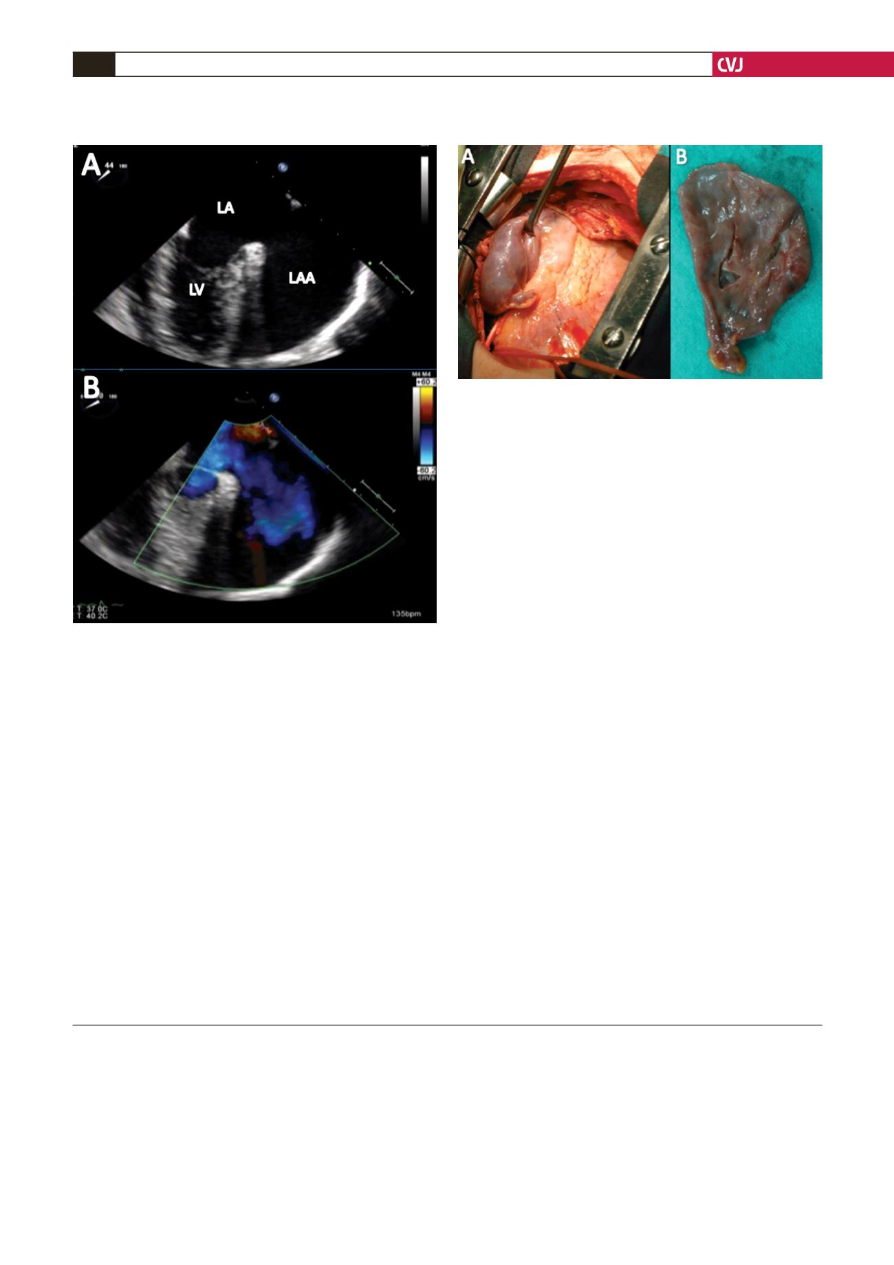
Fig. 3. A: Intra-operative image of the large LAA aneu-
rysm. B: Postoperative image of the resected material.
Fig. 2. Modified transoesophageal four-chamber, two-
dimensional (A), and colour flow (B) views showing a
huge LAA aneurysm with a broad left atrial connective
neck. There was no spontaneous echo-contrast or throm-
bus in the LAA and left atrium, and there was dynamic
blood flow within the aneurysm. LA: left atrium, LV: left
ventricle, LAA: left atrial appendage.


