


 19 / 102

CARDIOVASCULAR JOURNAL OF AFRICA • Volume 27, No 3, May/June 2016
AFRICA
141
techniques that require open-chest procedures and intubation
of vessels, offering no opportunity for time-course studies,
since they require time-consuming surgeries, with subsequent
euthanasia.
The Sonos 5500 echocardiographic system,
2-4,7
a universal type
of echocardiography instrument used in clinics, is commonly
equipped with a high-frequency transducer that can be used for
rodent studies. This study is the first report to use a standard
echocardiographic system (IE 33) to assess cardiac structure and
function in rats with myocardial hypertrophy. The IE 33 system
can clearly show parasternal long-axis and short-axis views,
apical four-chamber views of the LV, and the abdominal aorta in
rats without requiring an extra transducer. This reduces the cost
of investigation in small animals.
Concentric myocardial hypertrophy is a hallmark of chronic
pressure overload. Increased ventricular wall thickness induced
by overload pressure is initially beneficial to maintain normal
cardiac function. However, the heart could convert to heart
failure with LV dilatation if the hypertrophic stimulus is
maintained. In our study, significant increases in LVPW and
IVS with only small decreases in LVIDs were indicative of pure
concentric hypertrophy, which is often observed during the early
stages of pure pressure overload. Most previous studies have
shown late-stage chamber dilatation, signifying the end of the
compensatory response and the start of heart failure.
16
However,
in this study, even at six weeks post surgery, there was little
change in LVIDs, LVIDd, EDV and ESV, indicating chamber
dilatation and dysfunction in ventricular relaxation were not
present at this stage of hypertrophy.
FS and EF are commonly used parameters to evaluate systolic
function, which is determined by measuring LV end-diastolic
and end-systolic diameters with M-mode echocardiography.
Since increased afterload may depress stroke volume (SV) in
AAC rats, FS and EF were lower in the AAC rats compared
to sham rats from three to six weeks. Accordingly, significant
decreases in CO were observed in AAC rats, especially at the
four-week time point, since CO may be affected by HR and EF
(both reduced in the AAC groups).
In this study, we measured PFVA and E waves using
echocardiography to evaluate ventricular relaxation and diastolic
function. Significant decreases in PFVA and E-wave values in the
AAC rats suggested restrictive filling, which may result from the
combined effect of elevated ESV and impaired compliance due
to wall thickening and/or fibrosis.
17
Although E/A ratio is a marker for ventricular relaxation,
it could not be obtained in this study since the E and A waves
were fused in the rats due to their extremely fast heart rates.
To distinguish E and A waves, Kokubo
et al.
7
used ketamine
hydrochloride and xylazine anaesthesia in the animals in order
to decrease the HR to 70–80% of normal levels in conscious
animals, however control of HR is difficult.
In rats, the heart generally gains ~ 1 g in weight with each
2-mm increase in LV wall thickness. Progressive increases in
heart weight, HMI and LVMI, and increased cross-sectional
areas of the myocytes (determined using histological analysis)
were observed in the ACC rats. Elliott
et al.
3
found that
echocardiographic determination of LVm is relatively accurate,
yet highly overestimated in rats. As LVm increases, a greater
degree of LVm overestimation occurs. However, a strong
correlation between LVm determined using echocardiography
and the actual heart weights of sacrificed rats was observed in
the present study, indicating that the parameters measured using
echocardiography were closer to the actual values.
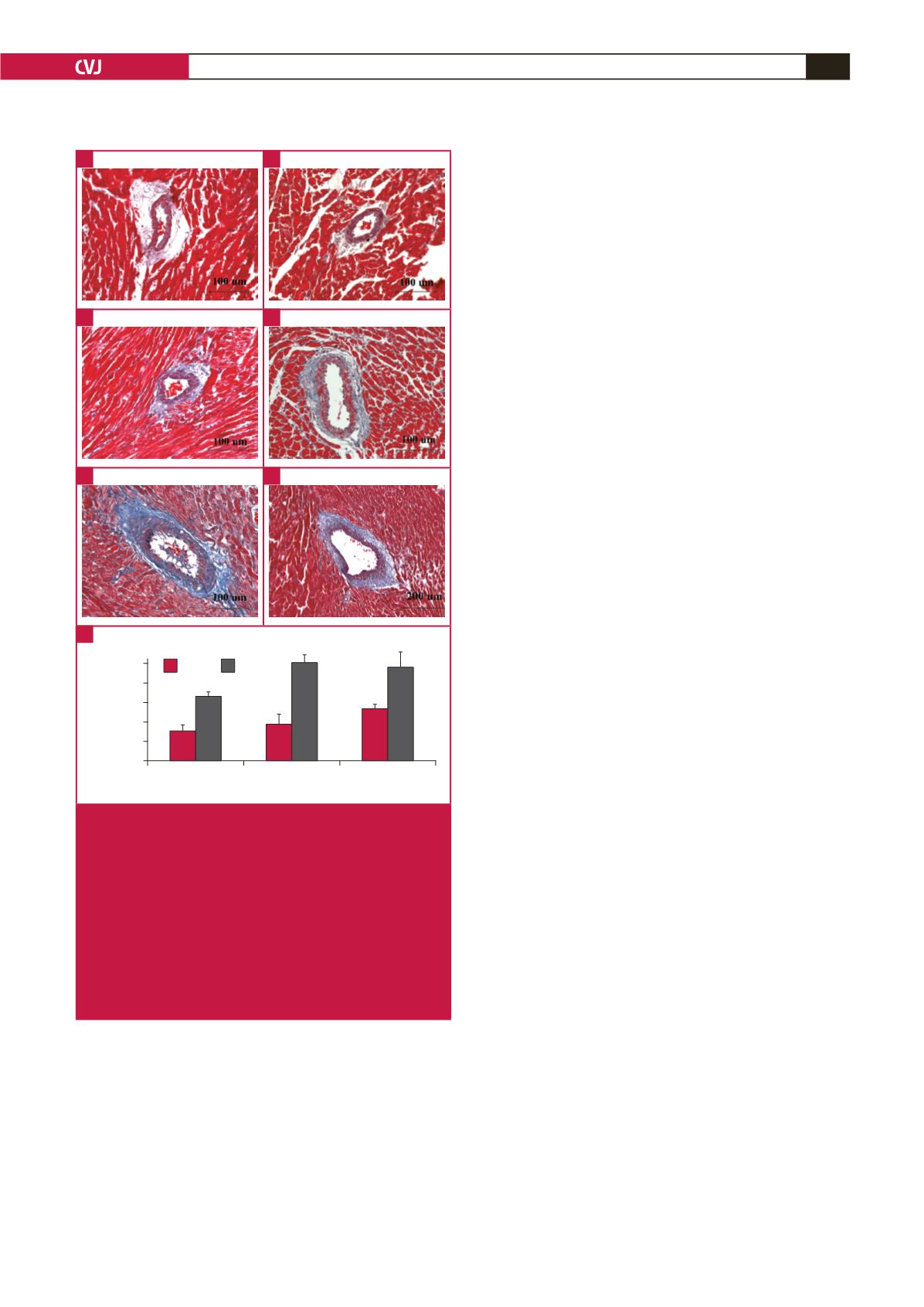
Fibrosis is a common response to pressure overload or
infarction, to overcome elevated ventricular wall stress. Excessive
fibrotic deposits around the small vessels may reduce oxygen
and nutrient exchange rates between the blood supply and the
surrounding myocardium. Additionally, extensive interstitial
fibrosis impedes myocardial relaxation, increasing stiffness in the
ventricular wall and reducing LV compliance.
3
4
6
Weeks
Perivascular fibrosis
(PVF/VA)
5
4
3
2
1
0
Sham AAC
**
**
*
Fig. 9.
Comparison of perivascular fibrosis between the sham
and AAC groups using Masson trichrome staining
(A–E) (
×
200), (E) (
×
100). Data are presented as mean
±
SEM. *
p
<
0.05 vs sham control, **
p
<
0.01 vs sham
control. A: sham rats at three weeks, B: sham rats at
four weeks, C: sham rats at six weeks, D: AAC rats at
three weeks, E: AAC rats at four weeks, F: AAC rats
at six weeks, G: quantitative analysis of perivascular
fibrosis between the sham and AAC groups. ANOVA
was performed to compare the AAC and sham groups
when the probability value was statistically significant.
An LSD
t
-test was applied for multiple comparisons.
A
C
E
G
B
D
F